

Long non-coding RNA MALAT1 enhances the apoptosis of cardiomyocytes through autophagy inhibition by regulating TSC2-mTOR signaling
- PMID: 31783925
- PMCID: PMC6883637
- DOI: 10.1186/s40659-019-0265-0
Long non-coding RNA MALAT1 enhances the apoptosis of cardiomyocytes through autophagy inhibition by regulating TSC2-mTOR signaling
Abstract
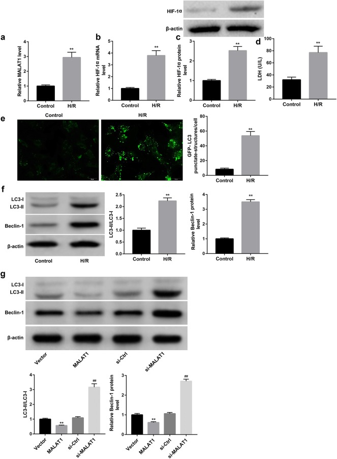
Background: Our previous study showed that knockdown of long noncoding RNA (lncRNA) metastasis-associated lung adenocarcinoma transcript 1 (MALAT1) attenuated myocardial apoptosis in mouse acute myocardial infarction (AMI). This study aims to explore whether MALAT1 enhanced cardiomyocyte apoptosis via autophagy regulation and the underlying mechanisms of MALAT1 regulating autophagy.
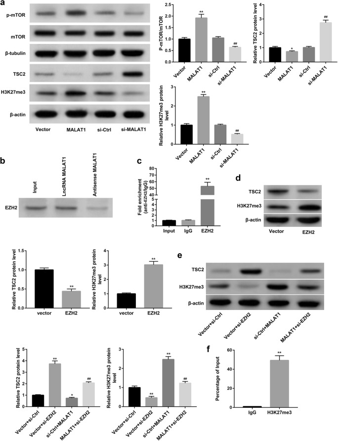
Methods: Cardiomyocytes were isolated from neonatal mice and then stimulated with hypoxia/reoxygenation (H/R) injury to mimic AMI. The autophagy level was assessed using GFP-LC3 immunofluorescence and western blot analysis of autophagy-related proteins. RNA pull-down and RNA immunoprecipitation (RIP) was performed to analyze the binding of MALAT1 and EZH2. Chromatin immunoprecipitation (ChIP) assay was performed to analyze the binding of TSC2 promoter and EZH2. The cell apoptosis was evaluated using TUNEL staining and western blot analysis of apoptosis-related proteins.
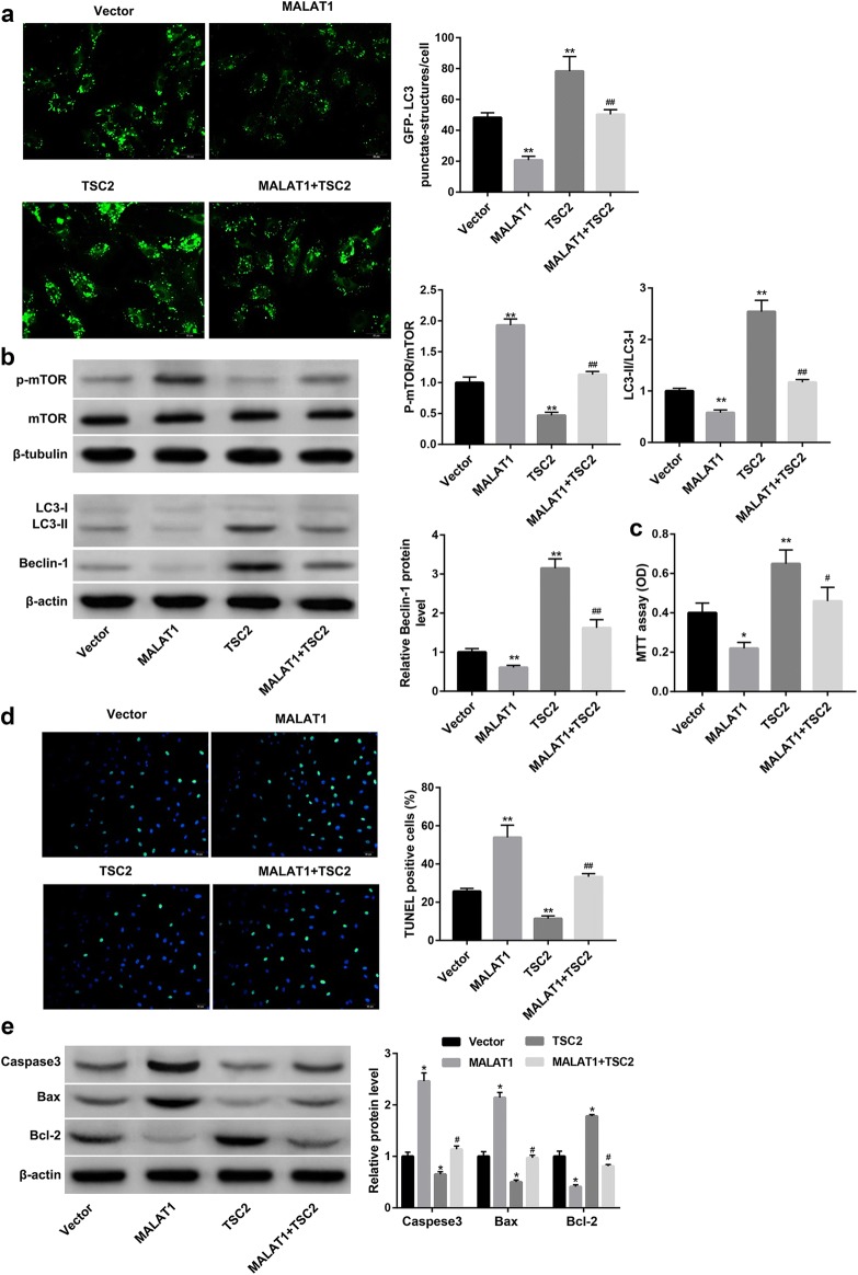
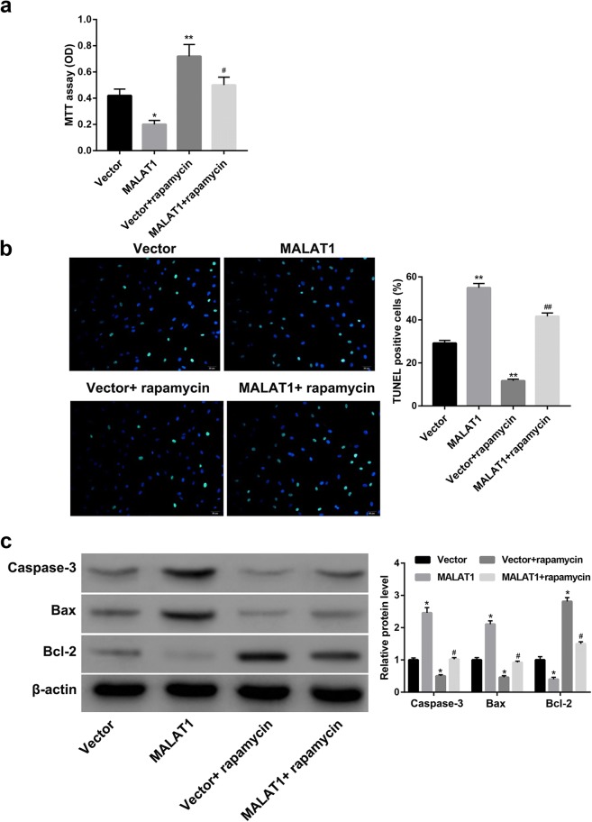
Results: H/R injury increased MALAT1 expression in cardiomyocytes. Furthermore, MALAT1 overexpression inhibited, whereas MALAT1 knockdown enhanced the autophagy of cardiomyocytes. Moreover, MALAT1 overexpression recruited EZH2 to TSC2 promoter regions to elevate H3K27me3 and epigenetically inhibited TSC2 transcription. Importantly, TSC2 overexpression suppressed mTOR signaling and then activated the autophagy. Further results showed that MALAT1 inhibited proliferation and enhanced apoptosis of cardiomyocytes through inhibiting TSC2 and autophagy.
Conclusion: These findings demonstrate that the increased MALAT1 expression induced by H/R injury enhances cardiomyocyte apoptosis through autophagy inhibition by regulating TSC2-mTOR signaling.
Keywords: Autophagy; Cardiomyocyte apoptosis; EZH2; MALAT1; TSC2-mTOR.
Conflict of interest statement
The authors declare that they have no competing interests.
Figures
Similar articles
-
Long noncoding RNA MALAT1 enhances the apoptosis of cardiomyocytes through autophagy modulation.Biochem Cell Biol. 2020 Apr;98(2):130-136. doi: 10.1139/bcb-2019-0062. Epub 2020 Jan 27. Biochem Cell Biol. 2020. PMID: 31985274
-
LncRNA MALAT1 protects cardiomyocytes from isoproterenol-induced apoptosis through sponging miR-558 to enhance ULK1-mediated protective autophagy.J Cell Physiol. 2019 Jul;234(7):10842-10854. doi: 10.1002/jcp.27925. Epub 2018 Dec 7. J Cell Physiol. 2019. PMID: 30536615
-
LncRNA MALAT1 Promotes Oxygen-Glucose Deprivation and Reoxygenation Induced Cardiomyocytes Injury Through Sponging miR-20b to Enhance beclin1-Mediated Autophagy.Cardiovasc Drugs Ther. 2019 Dec;33(6):675-686. doi: 10.1007/s10557-019-06902-z. Cardiovasc Drugs Ther. 2019. PMID: 31823095
-
Effect of Gpx3 gene silencing by siRNA on apoptosis and autophagy in chicken cardiomyocytes.J Cell Physiol. 2019 Jun;234(6):7828-7838. doi: 10.1002/jcp.27842. Epub 2018 Dec 4. J Cell Physiol. 2019. PMID: 30515791 Review.
-
Functions and regulatory mechanisms of metastasis-associated lung adenocarcinoma transcript 1.J Cell Physiol. 2018 Jan;234(1):134-151. doi: 10.1002/jcp.26759. Epub 2018 Aug 21. J Cell Physiol. 2018. PMID: 30132842 Review.
Cited by
-
Regulation of Pyroptosis by ncRNA: A Novel Research Direction.Front Cell Dev Biol. 2022 Mar 28;10:840576. doi: 10.3389/fcell.2022.840576. eCollection 2022. Front Cell Dev Biol. 2022. PMID: 35419365 Free PMC article. Review.
-
A Pan-Cancer Analysis of Transcriptome and Survival Reveals Prognostic Differentially Expressed LncRNAs and Predicts Novel Drugs for Glioblastoma Multiforme Therapy.Front Genet. 2021 Aug 24;12:723725. doi: 10.3389/fgene.2021.723725. eCollection 2021. Front Genet. 2021. PMID: 34759954 Free PMC article.
-
Using Machine Learning Methods in Identifying Genes Associated with COVID-19 in Cardiomyocytes and Cardiac Vascular Endothelial Cells.Life (Basel). 2023 Apr 14;13(4):1011. doi: 10.3390/life13041011. Life (Basel). 2023. PMID: 37109540 Free PMC article.
-
The role of CXCL8 in chronic nonhealing diabetic foot ulcers and phenotypic changes in fibroblasts: a molecular perspective.Mol Biol Rep. 2022 Feb;49(2):1565-1572. doi: 10.1007/s11033-022-07144-3. Epub 2022 Jan 19. Mol Biol Rep. 2022. PMID: 35044539 Review.
-
Promising roles of non-exosomal and exosomal non-coding RNAs in the regulatory mechanism and as diagnostic biomarkers in myocardial infarction.J Zhejiang Univ Sci B. 2023 Apr 15;24(4):281-300. doi: 10.1631/jzus.B2200459. J Zhejiang Univ Sci B. 2023. PMID: 37056205 Free PMC article. Review.
References
-
- Li M, Cheng W, Luo J, Hu X, Nie T, Lai H, et al. Loss of selenocysteine insertion sequence binding protein 2 suppresses the proliferation, migration/invasion and hormone secretion of human trophoblast cells via the PI3K/Akt and ERK signaling pathway. Placenta. 2017;55:81–89. doi: 10.1016/j.placenta.2017.05.007. - DOI - PubMed
MeSH terms
Substances
Grants and funding
LinkOut - more resources
Full Text Sources
Miscellaneous
