. 2020 Mar;97(3):609-614.
doi: 10.1016/j.kint.2019.08.029. Epub 2019 Sep 18.

A collagen-binding protein enables molecular imaging of kidney fibrosis in vivo

Affiliations

A collagen-binding protein enables molecular imaging of kidney fibrosis in vivo

Maike Baues et al. Kidney Int. 2020 Mar.

Abstract

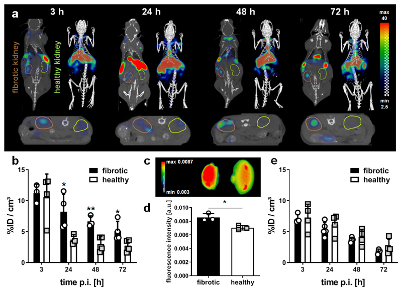
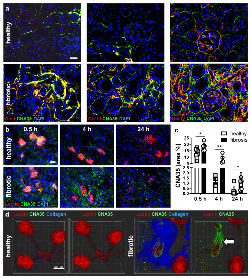
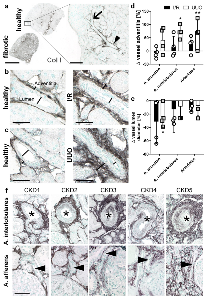
Pathological deposition of collagen is a hallmark of kidney fibrosis. To illustrate this process we employed multimodal optical imaging to visualize and quantify collagen deposition in murine models of kidney fibrosis (ischemia-reperfusion or unilateral ureteral obstruction) using the collagen-binding adhesion protein CNA35. For in vivo imaging, we used hybrid computed tomography-fluorescence molecular tomography and CNA35 labeled with the near-infrared fluorophore Cy7. Upon intravenous injection, CNA35-Cy7 accumulation was significantly higher in fibrotic compared to non-fibrotic kidneys. This difference was not detected for a non-specific scrambled version of CNA35-Cy7. Ex vivo, on kidney sections of mice and patients with renal fibrosis, CNA35-FITC co-localized with fibrotic collagen type I and III, but not with the basement membrane collagen type IV. Following intravenous injection, CNA35-FITC bound to both interstitial and perivascular fibrotic areas. In line with this perivascular accumulation, we observed significant perivascular fibrosis in the mouse models and in biopsy sections from patients with chronic kidney disease using computer-based morphometry quantification. Thus, molecular imaging of collagen using CNA35 enabled specific non-invasive quantification of kidney fibrosis. Collagen imaging revealed significant perivascular fibrosis as a consistent component next to the more commonly assessed interstitial fibrosis. Our results lay the basis for further probe and protocol optimization towards the clinical translation of molecular imaging of kidney fibrosis.

Keywords: chronic kidney disease (CKD); collagen; extracellular matrix; molecular imaging; non-invasive imaging; renal fibrosis.


Conflict of interest statement

Disclosure: No conflicts of interest to disclose.

Figures

Figure 1
Figure 1. In vivo and ex vivo optical imaging of collagen deposition kidneys with I/R-induced fibrosis using CNA35-Cy7.
(a) Coronal (left) and transversal (bottom) slices as well as 3D renderings (right) for hybrid computed tomography-fluorescence molecular tomography (CT-FMT) imaging of CNA35 biodistribution in a mouse with I/R-induced renal fibrosis at different time points after the i.v. injection of 1 nmol CNA35-Cy7. The fibrotic kidney is encircled in brown (left) and the healthy kidney in green (right) in the coronal view. (b) Quantification of the in vivo CT-FMT images shows prominent accumulation of CNA35-Cy7 in fibrotic kidneys at 24, 48 and 72 h after probe administration. (c, d) Ex vivo fluorescence reflectance imaging (FRI) at 72 h post injection confirms higher CNA35 accumulation in fibrotic kidneys (left). (e) Quantification of the kidney accumulation of scrambled CNA35-Cy7, demonstrating no specific accumulation in fibrotic vs. healthy kidneys. Values represent mean ± SD based on quantifying n=4 mice. *p<0.05, **p<0.01.
Figure 2
Figure 2. Microscopy imaging of collagen deposition in fibrotic kidneys using CNA35-FITC.
(a) Fluorescence microscopy analysis showing the co-localization of FITC-labeled CNA35 (green; upon incubation of tissue sections) with collagen fibers of type I, III and IV (all in red) on renal tissue sections from healthy mice and UUO mice. Nuclei are counterstained using DAPI (blue). Scale bar: 50 µm. (b) Representative immunofluorescence images showing the accumulation of FITC-labeled CNA35 (green) in healthy and fibrotic kidneys at 0.5, 4 and 24 h after i.v. injection. Perfused blood vessels are counterstained using rhodamine-lectin (red), cell nuclei using DAPI (blue). Scale bar: 50 µm. (c) Quantification of CNA35-FITC accumulation in fibrotic vs. healthy kidneys upon i.v. injection. (d) Two-photon laser scanning microscopy (TPLSM) indicated specific co-localization with collagen fibers (visualized using second harmonic generation) and much stronger enrichment of CNA35-FITC in fibrotic kidneys than in contralateral healthy kidneys, particularly around blood vessels (arrow). Scale bar: 50 µm. Values represent mean ± SD based on n=6 sections. *p<0.05, **p<0.01.
Figure 3
Figure 3. Perivascular fibrosis is a constant feature of renal fibrosis in animal models and patients.
(a) Localization and expression of collagen I in healthy and fibrotic renal tissue. The arrowhead points at the arteria arcuatae and the arrow indicates the arteria interlobulares. Scale bar: 2 mm. (b-e) Collagen type I is upregulated during renal fibrosis in both I/R (b) and UUO (c) not only in the interstitium, but also in the thickened adventitia of arterial vessels (d). Vessels showed a reduced vessel lumen diameter throughout the entire renal artery branches (e). Scale bar: 25 µm. (f) Enhanced collagen type III deposition in perivascular areas was also a common feature in human biopsies and increased with increasing CKD stage. The asterisks indicate the vessel lumen of arteria interlobulares and the arrowheads mark arteria afferens. Scale bar: 50 µm. Values represent mean ± SD based on quantifying n=4 mice per group. *p<0.05, **p<0.01 fibrotic vs. healthy kidney.

Comment in

References

    1. Djudjaj S, Boor P. Cellular and molecular mechanisms of kidney fibrosis. Mol Aspects Med. 2019;65:16–36. - PubMed
    1. Klinkhammer BM, Goldschmeding R, Floege J, et al. Treatment of Renal Fibrosis-Turning Challenges into Opportunities. Adv Chronic Kidney Dis. 2017;24:117–129. - PubMed
    1. Baues M, Dasgupta A, Ehling J, et al. Fibrosis imaging: Current concepts and future directions. Adv Drug Deliv Rev. 2017;121:9–26. - PMC - PubMed
    1. Sun Q, Baues M, Klinkhammer BM, et al. Elastin imaging enables noninvasive staging and treatment monitoring of kidney fibrosis. Sci Transl Med. 2019;11 - PMC - PubMed
    1. Krahn KN, Bouten CV, van Tuijl S, et al. Fluorescently labeled collagen binding proteins allow specific visualization of collagen in tissues and live cell culture. Anal Biochem. 2006;350:177–185. - PubMed

Publication types

LinkOut - more resources