. 2019 Nov 29;10(1):5470.
doi: 10.1038/s41467-019-13502-w.

Enzyme-mimetic self-catalyzed polymerization of polypeptide helices

Affiliations

Enzyme-mimetic self-catalyzed polymerization of polypeptide helices

Ziyuan Song et al. Nat Commun. .

Abstract

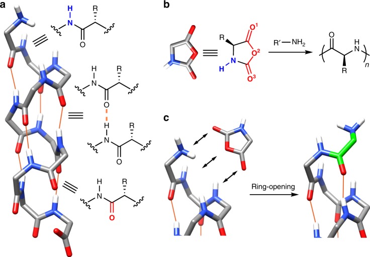
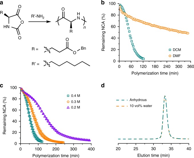
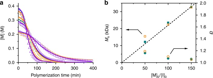
Enzymes provide optimal three-dimensional structures for substrate binding and the subsequent accelerated reaction. Such folding-dependent catalytic behaviors, however, are seldom mechanistically explored with reduced structural complexity. Here, we demonstrate that the α-helix, a much simpler structural motif of enzyme, can facilitate its own growth through the self-catalyzed polymerization of N-carboxyanhydride (NCA) in dichloromethane. The reversible binding between the N terminus of α-helical polypeptides and NCAs promotes rate acceleration of the subsequent ring-opening reaction. A two-stage, Michaelis-Menten-type kinetic model is proposed by considering the binding and reaction between the propagating helical chains and the monomers, and is successfully utilized to predict the molecular weights and molecular-weight distributions of the resulting polymers. This work elucidates the mechanism of helix-induced, enzyme-mimetic catalysis, emphasizes the importance of solvent choice in the discovery of new reaction type, and provides a route for rapid production of well-defined synthetic polypeptides by taking advantage of self-accelerated ring-opening polymerizations.


Conflict of interest statement

The authors declare no competing interests.

Figures

Fig. 1
Fig. 1
Schematic illustration of the catalytic power of an α-helix. a Illustration of possible binding sites at the termini of a free α-helical polypeptide. Intrahelical H-bonds are represented with orange lines. Side chains are omitted for simplicity. b Analysis of H-bond acceptors (red) and donors (blue) in an NCA molecule and its polymerization to synthesize polypeptides. c Proposed binding of NCA with the N terminus of an α-helix and subsequent ring-opening reaction for the growth of polypeptide. The newly formed residue from NCA is highlighted in green.
Fig. 2
Fig. 2
Polymerization kinetics of BLG-NCA. a Scheme showing the polymerization of BLG-NCA initiated by n-hexylamine. b Conversion of BLG-NCA in anhydrous DCM and DMF. [M]0 = 0.4 M, [M]0/[I]0 = 100. c Conversion of BLG-NCA in DCM at various [M]0. [M]0/[I]0 = 100. Error bars represent standard deviations from three independent measurements. d Normalized GPC-LS trace of obtained PBLG from polymerization of BLG-NCA in DCM under anhydrous conditions or in the presence of 10 vol% water. [M]0 = 0.4 M, [M]0/[I]0 = 100.
Fig. 3
Fig. 3
Reversible binding of polypeptide and NCA monomer. a, b Chemical structures of BLG-NCA, PBLG-NH2 a and PBLG-NHAc b. c STD NMR spectra of ELG-NCA in the presence of PBLG-NH2 or PBLG-NHAc. The STD signal of ring N‒H proton is highlighted with red arrows. The peak at 7.27 ppm is the irradiation peak since no background suppression was applied. d STD amplification factor of ring N‒H proton of ELG-NCA in the presence of PBLG-NH2 or PBLG-NHAc at various saturation times. e Snapshot of simulation trajectories showing the binding between PBLG-NH2 and BLG-NCA. The α-helical backbone is represented as a purple ribbon, the side chains of PBLG as sticks, and NCA as van der Waals spheres. f Closeup from the simulation trajectory at the N terminus of PBLG-NH2 to reveal the H-bonding interactions (highlighted with the orange dotted lines). Side chains are omitted for simplicity. g Time evolution of the H bonds formed between BLG-NCA (O1) and PBLG-NH2 (amide N–H from third residue). Each line represents one independent, 2-μs long MD simulation. The red arrow indicates the transfer of NCA from C terminus to N terminus in the third simulation run. h PMF profile for the reversible binding of PBLG-NH2 and BLG-NCA. Error bars correspond to estimated standard deviations from four independent walkers of the eABF algorithm.
Fig. 4
Fig. 4
Simulations with adsorption-incorporated, two-stage kinetic model. a Plot of the monomer concentration vs. time for test cases with s = 10, [M]0/[I]0 = 100, k1 = 0.02 M−1 s−1, kon = 10 M−1 s−1, koff = 2 s−1, kr = 0.2 s−1, and at selected values of [M]0 = 0.05, 0.1, 0.2, or 0.4 M. b Predicted MWD profiles based on the kinetic profiles in a. c Calculated DP and Đ at various [M]0.
Fig. 5
Fig. 5
Kinetic modeling of polymerization of NCA in DCM. a Polymerization kinetics in Fig. 2c were fit with the adsorption-incorporated kinetic model. b Comparison of experimental results obtained from GPC analysis (solid symbols) and predicted results from modeling (open symbols) with various [M]0/[I]0 ratios at [M]0 = 0.4 M. The dashed line represents designed MW by [M]0/[I]0 ratio.

References

    1. Cannon WR, Singleton SF, Benkovic SJ. A perspective on biological catalysis. Nat. Struct. Biol. 1996;3:821–833. doi: 10.1038/nsb1096-821. - DOI - PubMed
    1. Benkovic SJ, Hammes-Schiffer S. A perspective on enzyme catalysis. Science. 2003;301:1196–1202. doi: 10.1126/science.1085515. - DOI - PubMed
    1. Wolfenden R, Snider MJ. The depth of chemical time and the power of enzymes as catalysts. Acc. Chem. Res. 2001;34:938–945. doi: 10.1021/ar000058i. - DOI - PubMed
    1. Purich DL. Enzyme Kinetics: Catalysis and Control. Amsterdam: Elsevier Science; 2010.
    1. Bartlett GJ, Porter CT, Borkakoti N, Thornton JM. Analysis of catalytic residues in enzyme active sites. J. Mol. Biol. 2002;324:105–121. doi: 10.1016/S0022-2836(02)01036-7. - DOI - PubMed

Publication types