. 2019 Nov 29;6(1):295.
doi: 10.1038/s41597-019-0303-3.

A manually denoised audio-visual movie watching fMRI dataset for the studyforrest project

Affiliations

A manually denoised audio-visual movie watching fMRI dataset for the studyforrest project

Xingyu Liu et al. Sci Data. .

Abstract

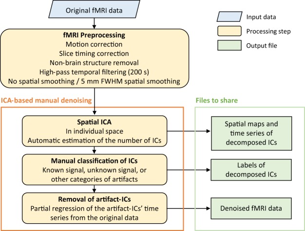
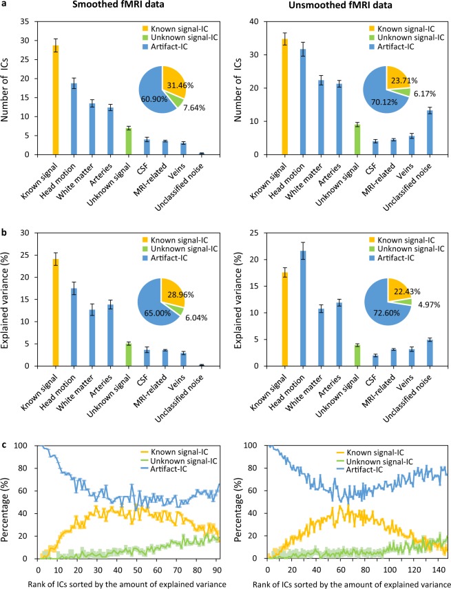
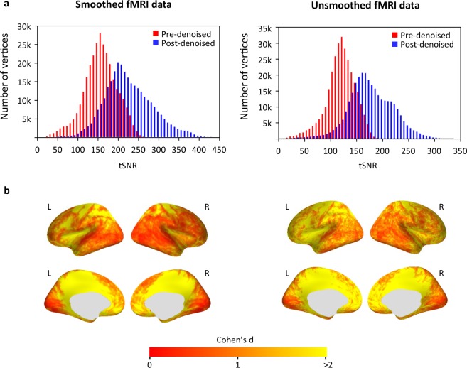
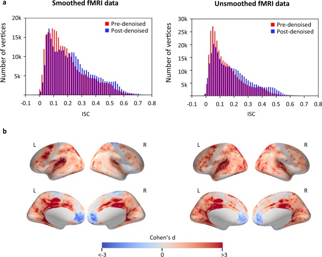
The data presented here are related to the studyforrest project that uses the movie 'Forrest Gump' to map brain functions in a real-life context using functional magnetic resonance imaging (fMRI). However, neural-related fMRI signals are often small and confounded by various noise sources (i.e., artifacts) that makes searching for the signals induced by specific cognitive processes significantly challenging. To make neural-related signals stand out from the noise, the audio-visual movie watching fMRI dataset from the project was denoised by a combination of spatial independent component analysis and manual identification of signals or noise. Here, both the denoised data and the labeled decomposed components are shared to facilitate further study. Compared with the original data, the denoised data showed a substantial improvement in the temporal signal-to-noise ratio and provided a higher sensitivity in subsequent analyses such as in an inter-subject correlation analysis.


Conflict of interest statement

The authors declare no competing interests.

Figures

Fig. 1
Fig. 1
A schematic overview of the four-step denoising workflow. Preprocessing was performed on the original fMRI data and included motion correction, slice timing correction, non-brain structure removal, high-pass filtering (200 s cut-off); all steps were performed without or with spatial smoothing (i.e., FWHM = 0 mm or FWHM = 5 mm). A spatial ICA was next run on the preprocessed data in individual space, producing spatial maps and time series files of the decomposed ICs for each run and each participant. All decomposed ICs were then manually classified into either a known signal, unknown signal, or different categories of artifacts. Finally, all the ICs classified as artifacts were filtered out, producing the final denoised fMRI data. (fMRI: functional magnetic resonance imaging, FWHM: full width half maximum, IC: independent component, ICA: independent component analysis).
Fig. 2
Fig. 2
Artifact ICs accounted for a large proportion of ICs and variances in both smoothed (left column) and unsmoothed (right column) fMRI data. (a) The number of ICs classified into different categories is shown. The number of ICs was averaged across all runs and participants. (b) The explained variance of ICs in different categories is shown. The explained variance of all ICs in each category was totaled at each run, then averaged across all runs and participants. (c) The proportions of known signal-ICs, unknown signal-ICs, and artifact-ICs for each rank are shown. ICs from each run were ranked according to the amount of variance they explained, and the proportions of the three superordinate IC categories were summarized across all runs and participants for each rank. The error bars in (a) and (b) and shaded areas in (c) denote the standard error (SEM) of participants (n = 15). (fMRI: functional magnetic resonance imaging, IC: independent component, CSF: cerebral spinal fluid).
Fig. 3
Fig. 3
Denoised fMRI data showed higher tSNR than the original data (left: smoothed fMRI data; right: unsmoothed fMRI data). (a) Histograms of the tSNR values across all vertices on the fsaverage surface are shown. The tSNR was calculated for each vertex and averaged across all runs and participants. (b) The Cohen’s d effect size of the tSNR change is displayed on the fsaverage surface. Cohen’s d was calculated as the mean differences between the tSNR from pre- and post-denoised fMRI data (n = 15 participants), divided by the pooled standard deviation. (fMRI: functional magnetic resonance imaging, tSNR: temporal signal-to-noise ratio, L: left hemisphere, R: right hemisphere).
Fig. 4
Fig. 4
Denoised fMRI data showed higher ISC than the original data in movie watching-related brain regions (left: smoothed fMRI data; right: unsmoothed fMRI data). (a) Histograms of ISCs across all vertices on the fsaverage surface are shown. The ISC value was averaged across all runs and participants. (b) The Cohen’s d effect size of ISC changes at each vertex on the fsaverage surface is shown. The ISC coefficients were first converted into z-values using a Fisher’s r-to-z transformation. Next, the Cohen’s d value was calculated as the differences in means between the z-values from pre- and post-denoised fMRI data (n = 8 runs), divided by the pooled standard deviation. (fMRI: functional magnetic resonance imaging, ISC: inter-subject correlation, L: left hemisphere, R: right hemisphere).

Dataset use reported in

  • doi: 10.1038/sdata.2014.3
  • doi: 10.1038/sdata.2016.92

References

    1. Spiers HJ, Maguire EA. Decoding human brain activity during real-world experiences. Trends Cogn. Sci. 2007;11:356–365. doi: 10.1016/j.tics.2007.06.002. - DOI - PubMed
    1. Hasson U, et al. Neurocinematics: The neuroscience of film. Projections. 2008;2:1–26. doi: 10.3167/proj.2008.020102. - DOI
    1. Bartels A, Zeki S. The chronoarchitecture of the human brain—natural viewing conditions reveal a time-based anatomy of the brain. NeuroImage. 2004;22:419–433. doi: 10.1016/j.neuroimage.2004.01.007. - DOI - PubMed
    1. Hasson U, Malach R, Heeger DJ. Reliability of cortical activity during natural stimulation. Trends Cogn. Sci. 2010;14:40–48. doi: 10.1016/j.tics.2009.10.011. - DOI - PMC - PubMed
    1. Vanderwal T, Kelly C, Eilbott J, Mayes LC, Castellanos FX. Inscapes: A movie paradigm to improve compliance in functional magnetic resonance imaging. NeuroImage. 2015;122:222–232. doi: 10.1016/j.neuroimage.2015.07.069. - DOI - PMC - PubMed

Publication types