

Highly cooperative fluorescence switching of self-assembled squaraine dye at tunable threshold temperatures using thermosensitive nanovesicles for optical sensing and imaging
- PMID: 31784685
- PMCID: PMC6884458
- DOI: 10.1038/s41598-019-54418-1
Highly cooperative fluorescence switching of self-assembled squaraine dye at tunable threshold temperatures using thermosensitive nanovesicles for optical sensing and imaging
Abstract
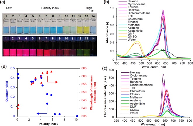
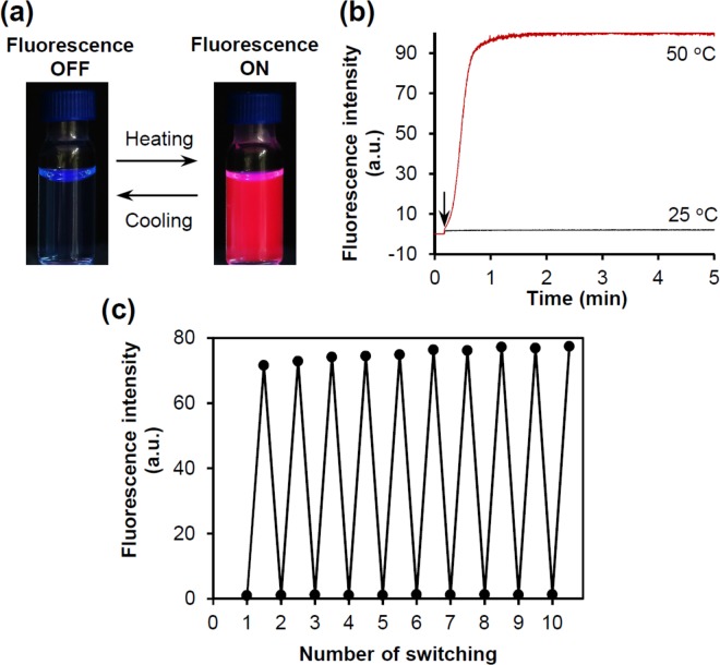
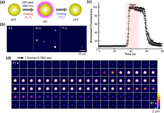
Thermosensitive fluorescent dyes can convert thermal signals into optical signals as a molecular nanoprobe. These nanoprobes are playing an increasingly important part in optical temperature sensing and imaging at the nano- and microscale. However, the ability of a fluorescent dye itself has sensitivity and accuracy limitations. Here we present a molecular strategy based on self-assembly to overcome such limitations. We found that thermosensitive nanovesicles composed of lipids and a unique fluorescent dye exhibit fluorescence switching characteristics at a threshold temperature. The switch is rapid and reversible and has a high signal to background ratio (>60), and is also highly sensitive to temperature (10-22%/°C) around the threshold value. Furthermore, the threshold temperature at which fluorescence switching is induced, can be tuned according to the phase transition temperature of the lipid bilayer membrane forming the nanovesicles. Spectroscopic analysis indicated that the fluorescence switching is induced by the aggregation-caused quenching and disaggregation-induced emission of the fluorescent dye in a cooperative response to the thermotropic phase transition of the membrane. This mechanism presents a useful approach for chemical and material design to develop fluorescent nanomaterials with superior fluorescence sensitivity to thermal signals for optical temperature sensing and imaging at the nano- and microscales.
Conflict of interest statement
The authors declare no competing interests.
Figures


Similar articles
-
New fluorescent labels with tunable hydrophilicity for the rational design of bright optical probes for molecular imaging.Bioconjug Chem. 2013 Jul 17;24(7):1174-85. doi: 10.1021/bc4000349. Epub 2013 Jun 26. Bioconjug Chem. 2013. PMID: 23758616
-
Media Dependent Switching of Selectivity and Continuous near Infrared Turn-on Fluorescence Response through Cascade Interactions from Noncovalent to Covalent Binding for Detection of Serum Albumin in Living Cells.ACS Appl Mater Interfaces. 2018 Dec 26;10(51):44336-44343. doi: 10.1021/acsami.8b19768. Epub 2018 Dec 13. ACS Appl Mater Interfaces. 2018. PMID: 30514088
-
Collective fluorescence switching of counterion-assembled dyes in polymer nanoparticles.Nat Commun. 2014 Jun 9;5:4089. doi: 10.1038/ncomms5089. Nat Commun. 2014. PMID: 24909912
-
The problem of self-calibration of fluorescence signal in microscale sensor systems.Lab Chip. 2005 Nov;5(11):1210-23. doi: 10.1039/b507447a. Epub 2005 Sep 27. Lab Chip. 2005. PMID: 16234943 Review.
-
Ratiometric fluorescent nanoprobes for visual detection: Design principles and recent advances - A review.Anal Chim Acta. 2019 Nov 4;1079:30-58. doi: 10.1016/j.aca.2019.06.035. Epub 2019 Jun 17. Anal Chim Acta. 2019. PMID: 31387719 Review.
Cited by
-
A rapid and highly sensitive biomarker detection platform based on a temperature-responsive liposome-linked immunosorbent assay.Sci Rep. 2020 Oct 22;10(1):18086. doi: 10.1038/s41598-020-75011-x. Sci Rep. 2020. PMID: 33093468 Free PMC article.
-
Temperature-Responsive Liposome-Linked Immunosorbent Assay for the Rapid Detection of SARS-CoV-2 Using Immunoliposomes.ACS Omega. 2022 Jul 21;7(30):26936-26944. doi: 10.1021/acsomega.2c03597. eCollection 2022 Aug 2. ACS Omega. 2022. PMID: 35915635 Free PMC article.
References
-
- Arai, S. & Suzuki, M. Nanosized optical thermometers. In Smart nanoparticles for biomedicine (ed. Ciofani, G.) Chapter 14, 199–217 (Elsevier, 2018).
Publication types
LinkOut - more resources
Full Text Sources
Other Literature Sources
