. 2020 May;67(3):1129-1140.
doi: 10.1111/tbed.13439. Epub 2019 Dec 13.

A molecular epidemiological investigation of PEDV in China: Characterization of co-infection and genetic diversity of S1-based genes

Affiliations

A molecular epidemiological investigation of PEDV in China: Characterization of co-infection and genetic diversity of S1-based genes

Mingjun Su et al. Transbound Emerg Dis. 2020 May.

Erratum in

  • Corrigendum.
    [No authors listed] [No authors listed] Transbound Emerg Dis. 2021 Jul;68(4):2634-2635. doi: 10.1111/tbed.14152. Epub 2021 Jun 5. Transbound Emerg Dis. 2021. PMID: 34089234 Free PMC article. No abstract available.

Abstract

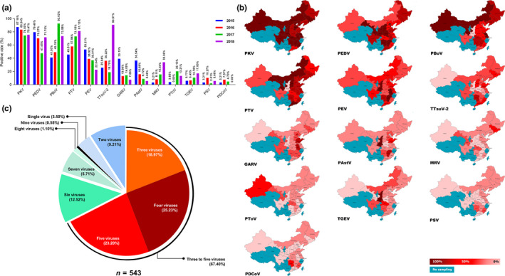
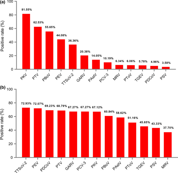
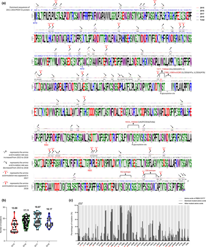
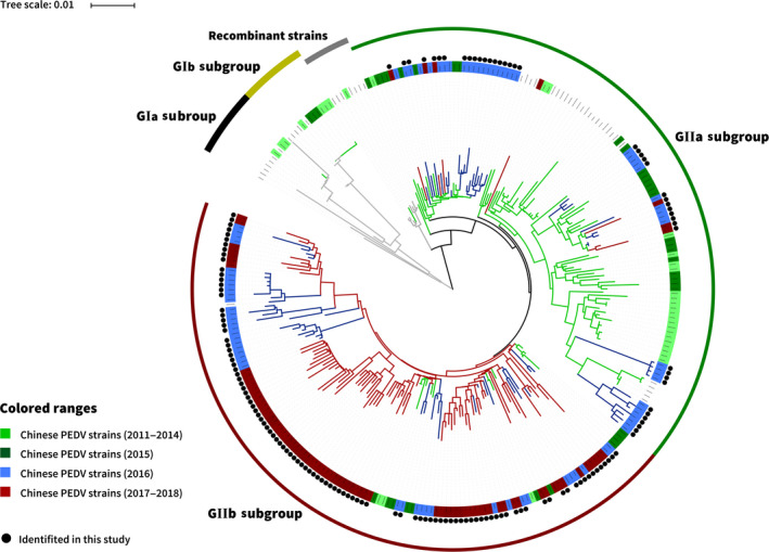
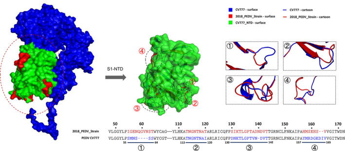
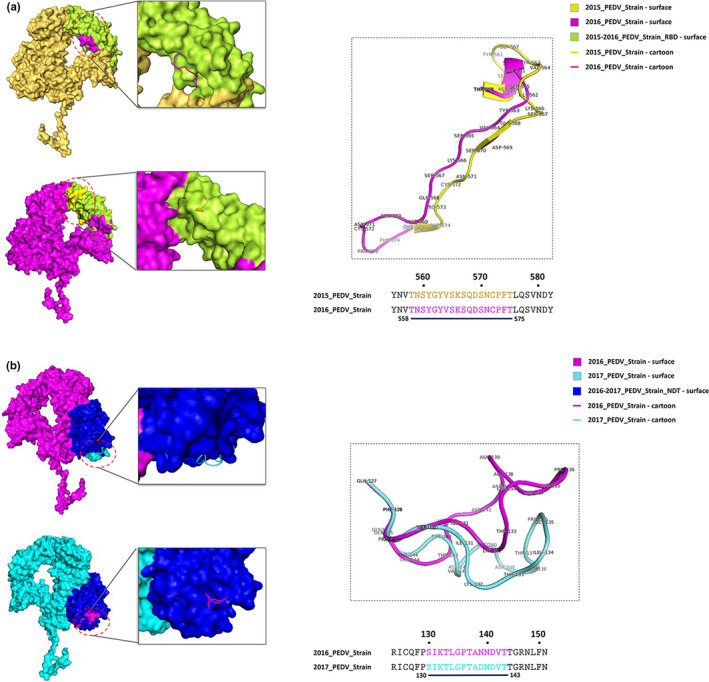
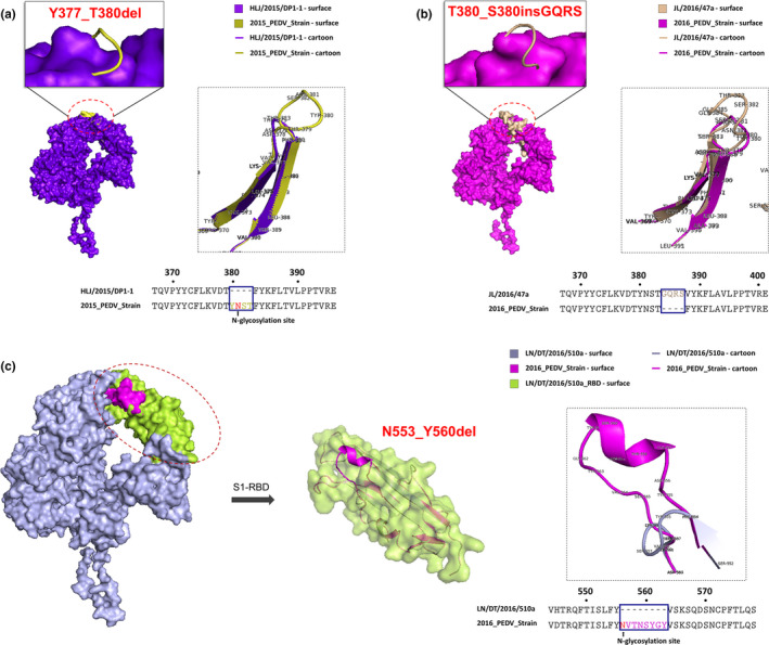
Porcine epidemic diarrhoea virus (PEDV) is an emerging and re-emerging epizootic virus of swine that causes substantial economic losses to the pig industry in China and other countries. The variations in the virus, and its co-infections with other enteric viruses, have contributed to the poor control of PEDV infection. In the current study, a broad epidemiological investigation of PEDV was carried out in 22 provinces or municipalities of China during 2015-2018. The enteric viruses causing co-infection with PEDV and the genetic diversity of the PEDV S1 gene were also analysed. The results indicated that, of the 543 diarrhoea samples, 66.85% (363/543) were positive for PEDV, and co-infection rates of PEDV with 13 enteric viruses ranged from 3.58% (13/363) to 81.55% (296/363). Among these enteric viruses, the signs of diarrhoea induced by PEDV were potentially associated with co-infections with porcine enterovirus 9/10 (PEV) and torque teno sus virus 2 (TTSuV-2) (p < .05). The 147 PEDV strains identified in our study belong to Chinese pandemic strains and exhibited genetic diversity. The virulence-determining S1 proteins of PEDV pandemic strains were undergoing amino acid mutations, in which S58_S58insQGVN-N135dup-D158_I159del-like mutations were common patterns (97.28%, 143/147). When compared with 2011-2014 PEDV strains, the amino acid mutations of PEDV pandemic strains were mainly located in the N-terminal domain of S1 (S1-NTD), and 21 novel mutations occurred in 2017 and 2018. Furthermore, protein homology modelling showed that the mutations in pattern of insertion and deletion mutations of the S1 protein of PEDV pandemic strains may have caused structural changes on the surface of the S1 protein. These data provide a better understanding of the co-infection and genetic evolution of PEDV in China.

Keywords: PEDV; S1 gene; co-infection; mutation.


Conflict of interest statement

The authors declare no conflict of interest.

Figures

Figure 1
Figure 1
The positive rate and geographic distribution of the selected enteric viruses in the diarrhoea samples during 2015–2018. (a) The positive rate of the selected 13 enteric viruses in the 543 diarrhoea samples during 2015–2018. (b) The geographic distribution of the selected 13 enteric viruses in the 543 diarrhoea samples during 2015–2018. (c) The mix‐infection patterns of the selected 13 enteric viruses in the 543 diarrhoea samples during 2015–2018 [Colour figure can be viewed at wileyonlinelibrary.com]
Figure 2
Figure 2
The co‐infection of PEDV with the selected enteric viruses in the diarrhoea samples during 2015–2018. (a) The co‐infection rate of the selected 13 enteric viruses in 363 PEDV‐positive samples. (b) The co‐infection rate of PEDV in the 13 enteric viruses positive samples [Colour figure can be viewed at wileyonlinelibrary.com]
Figure 3
Figure 3
Sequence analysis of S1 proteins of the PEDV strains identified in our study. (a) Divergence analysis of S1 proteins of PEDV strains identified in our study during 2015–2018. (b) The comparison of amino acid mutations of S1 proteins of PEDV strains identified in our study during 2015–2018 with PEDV CV777 strain. (c) The comparison of amino acid mutation positions of S1 proteins of PEDV strains identified in our study during 2015–2018 with PEDV CV777 strain [Colour figure can be viewed at wileyonlinelibrary.com]
Figure 4
Figure 4
The S1‐based phylogenetic analysis of the PEDV strains identified in our study. Black circle diagram represents the 147 PEDV strains identified in this study [Colour figure can be viewed at wileyonlinelibrary.com]
Figure 5
Figure 5
Comparative analysis of the predicted S1 protein modelling between PEDV CV777 strain and 2018_PEDV‐strain. The S1 protein modelling of PEDV CV777 strain was shown as surface and cartoon by blue. The S1 protein modelling of 2018_PEDV‐strian was shown as surface and cartoon by red. The mutant amino acid residues of S1 protein of 2018_PEDV‐strain were shown as surface by red. The S1‐NTD of PEDV CV777 strain was shown as surface by green [Colour figure can be viewed at wileyonlinelibrary.com]
Figure 6
Figure 6
Comparative analysis of the predicted S1 protein modelling among 2015–2018 PEDV strains. (a) Comparative analysis of the predicted S1 protein modelling between 2015_PEDV‐strain and 2016_PEDV‐strain. (b) The compared S1 modelling of the 2016_PEDV‐strain and 2017_PEDV‐strain. The 2015_PEDV‐strain was shown as surface and cartoon by yellow; 2016_PEDV‐strain was shown as surface and cartoon by pink; 2017_PEDV‐strain was shown as surface and cartoon by cyan. The S1‐NDT of 2016‐2017_PEDV‐strain was shown as surface by blue. The S1‐RBD 2015‐2016_PEDV‐strain was shown as surface by limon [Colour figure can be viewed at wileyonlinelibrary.com]
Figure 7
Figure 7
Comparative analysis of the predicted S1 protein modelling of PEDV HLJ/2015/DP1‐1, JL/2016/47a and LN/DT/2016/510a strains. (a) The compared S1 protein modelling between PEDV HLJ/2015/DP1‐1 strain and 2015_PEDV‐strain. (b) The compared S1 protein modelling between PEDV JL/2016/47a strain and 2016_PEDV‐strain. (c) The compared S1 protein modelling between PEDV LN/DT/2016/510a strain and 2016_PEDV‐strain. The HLJ/2015/DP‐1‐1 strain was shown as surface and cartoon by purpleblue. The 2015_PEDV‐strain was shown as cartoon by yellow. The JL/2016/47a strain was shown as surface and cartoon by wheat. The LN/DT/2016/510a strain was shown as surface and cartoon by lightblue. The 2016_PEDV‐strain was shown as surface and cartoon by pink. The S1‐RBD of LN/DT/2016/510a strain was shown as surface by limon [Colour figure can be viewed at wileyonlinelibrary.com]

References

    1. Aydin, H. , Al‐Khooly, D. , & Lee, J. E. (2014). Influence of hydrophobic and electrostatic residues on SARS‐coronavirus S2 protein stability: Insights into mechanisms of general viral fusion and inhibitor design. Protein Science, 23(5), 603–617. 10.1002/pro.2442 - DOI - PMC - PubMed
    1. Biasini, M. , Bienert, S. , Waterhouse, A. , Arnold, K. , Studer, G. , Schmidt, T. , Schwede, T. (2014). SWISS‐MODEL: Modeling protein tertiary and quaternary structure using evolutionary information. Nucleic Acids Research, 42, W252–W258. 10.1007/978-1-4939-8736-8_17 - DOI - PMC - PubMed
    1. Chen, P. , Wang, K. , Hou, Y. , Li, H. , Li, X. , Yu, L. , … Zhou, Y. (2019). Genetic evolution analysis and pathogenicity assessment of porcine epidemic diarrhea virus strains circulating in part of China during 2011–2017. Infection, Genetics and Evolution, 69, 153–165. 10.1016/j.meegid.2019.01.022 - DOI - PMC - PubMed
    1. Chen, Q. , Wang, L. , Zheng, Y. , Zhang, J. , Guo, B. , Yoon, K. J. , Li, G. (2018). Metagenomic analysis of the RNA fraction of the fecal virome indicates high diversity in pigs infected by porcine endemic diarrhea virus in the United States. Virology Journal, 15(1), 95. 10.1186/s12985-018-1001-z - DOI - PMC - PubMed
    1. Chu, D. K. , Poon, L. L. , Guan, Y. , & Peiris, J. S. (2008). Novel astroviruses in insectivorous bats. Journal of Virology, 82, 9107–9114. 10.1128/JVI.00857-08 - DOI - PMC - PubMed

MeSH terms

Associated data

LinkOut - more resources