Review
. 2020;181(1):11-23.
doi: 10.1159/000504847. Epub 2019 Nov 29.

The Cellular Functions of Eosinophils: Collegium Internationale Allergologicum (CIA) Update 2020

Affiliations
Review

The Cellular Functions of Eosinophils: Collegium Internationale Allergologicum (CIA) Update 2020

Hans-Uwe Simon et al. Int Arch Allergy Immunol. 2020.

Abstract

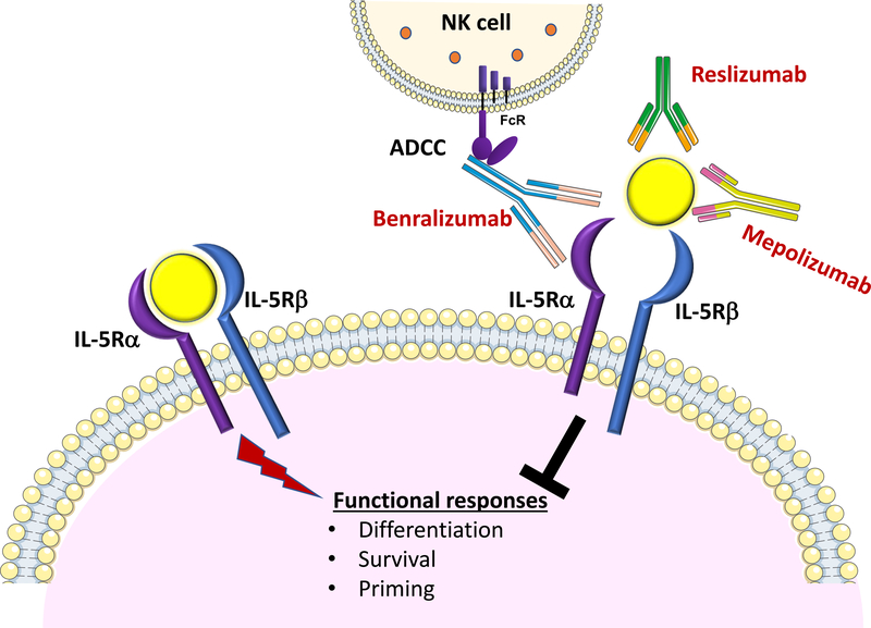
Eosinophils and their secretory mediators play an important role in the pathogenesis of infectious and inflammatory disorders. Although eosinophils are largely evolutionally conserved, their physiologic functions are not well understood. Given the availability of new eosinophil-targeted depletion therapies, there has been a renewed interest in understanding eosinophil biology as these strategies may result in secondary disorders when applied over long periods of time. Recent data suggest that eosinophils are not only involved in immunological effector functions but also carry out tissue protective and immunoregulatory functions that actively contribute to the maintenance of homeostasis. Prolonged eosinophil depletion may therefore result in the development of secondary disorders. Here, we review recent literature pointing to important roles for eosinophils in promoting immune defense, antibody production, activation of adipose tissue, and tissue remodeling and fibrosis. We also reflect on patient data from clinical trials that feature anti-eosinophil therapeutics.

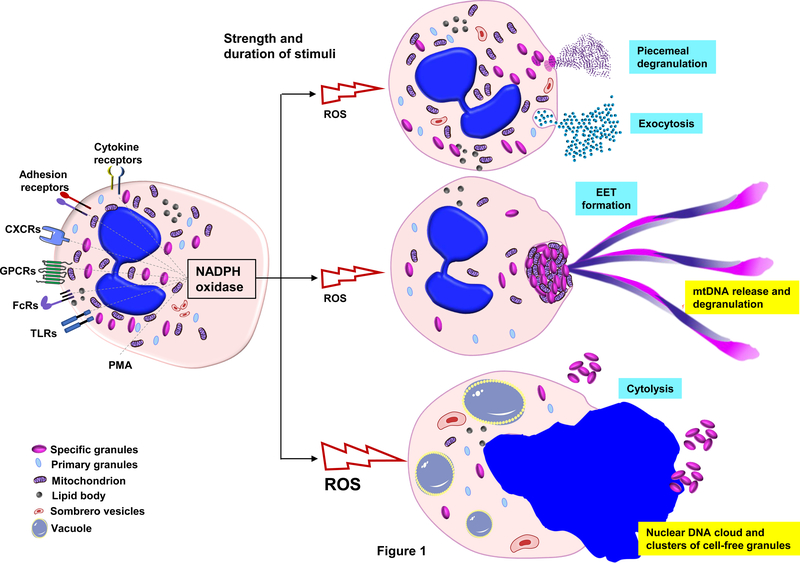
Keywords: Asthma; Eosinophil cytolysis; Eosinophil degranulation; Eosinophil extracellular traps; Eosinophils; Hypereosinophilic syndromes; Inflammation; Interleukin-5; M2 macrophages; Major basic protein; Mast cells; Targeted therapy.


Figures

Figure 1.
Figure 1.. Mediator release from eosinophils: degranulation, extracellular traps, and cytolysis
Schematic representation of functional responses leading to release of secretory mediators. In response physiologic stimuli, eosinophils respond by assembling NADPH oxidase and releasing reactive oxygen species (ROS). Depending on the source, strength and duration of the initial stimulus, eosinophils can degranulate, generate extracellular traps, or undergo cytolysis. Degranulation most typically occurs via by piecemeal degranulation or exocytosis of mediators from the cytoplasmic granules as shown. Eosinophils can also form extracellular traps (EETs), which consist of a mitochondrial (mt)DNA scaffold and cationic granule proteins. Cell activation above a certain threshold can lead to a non-apoptotic form of cell death, designated as cytolysis. During cytolysis cells form large vacuoles. The vacuoles, plasma and nuclear membranes ultimately disintegrate, leading to release of the nuclear DNA as a virtual “cloud”. Release of extracellular clusters of cell-free granules are typically observed as a result of cytolysis.
Figure 2.
Figure 2.. Eosinophil extracellular traps (EETs) entrap and kill bacteria
Confocal microscopy. E. coli triggers release of mtDNA and cationic granule proteins from mouse eosinophils. Mouse eosinophils seeded on glass coverslips were primed with GM-CSF for 20 min and subsequently co-cultured with GFP-labelled E. coli (ratio of 1:10) for 15 min. In the final 5 min of the stimulation period, cells were labelled with 5 μM MitoSOX Red to stain the extracellular DNA. Cells were then fixed with 4% paraformaldehyde and the nucleus stained with 1 μM Hoechst 33342. The image was acquired by LSM 700 (Carl Zeiss Micro Imaging, Jena, Germany) using an 63x /1.40 Oil DIC objective. Bars, 10 μm.
Figure 3.
Figure 3.. Eosinophil - mast cell interactions
Schematic representation of interactions between eosinophils and mast cells. Eosinophils release pre-formed mediators including stem cell factor (SCF), nerve growth factor (NGF), and leukotrienes C4 and E4 (LTC4/E4); these mediators elicit a functional response through activation of specific receptors on mast cells, including c-KIT receptor (CD117), tropomyosin-receptor kinase A (Trak A), and leukotriene receptors (LTC4R and LTE4R). Mast cell degranulation promoted by allergens via cross-linking of high-affinity immunoglobulin E receptors (FcεRI) results in release of histamine that can acts on eosinophils through the histamine receptor 4 (HR4). Activated mast cells also regulate eosinophil function via the activation of cytokine receptors, leukotriene receptors (LTB4R and LTC4R), prostaglandin D2 (PGD2) receptor (CRTH2), chemokine receptors (CXCRs) and other G-protein coupled receptors (GPCRs). Mast cells and eosinophils also maintain inhibitory immunoreceptor tyrosine-based inhibition motif (ITIM)-bearing receptors (IRs) and Siglecs, both of which serve to downregulate immune responses (128).
Figure 4.
Figure 4.. Eosinophils regulate the metabolism of adipose tissue
Schematic representation of the potential role of eosinophils in adipocyte metabolism. The current view is that IL-33 from resident mesenchyme-derived stromal cells in white adipose tissue activates resident type 2 innate lymphoid cell (ILC2) to produce IL-5 (129); this cytokine mediator activates eosinophils, which facilitates both chemoattraction and prolonged survival. Activated eosinophils release IL-4, which, together with ILC2, contribute IL-13 and convert tissue resident macrophages to alternatively activated macrophages (AAMs) capable of producing and releasing the norepinephrine required for adipose browning (130), although the parts of the pathway linking AAMs and norepinephrine to the browning process have recently been questioned (131). Other data suggest that eosinophils may also interact with adipocytes directly by releasing unknown eosinokines (noted in the figure with “?” inferring soluble factors from eosinophils that initiate the browning process). In addition, adipocytes can release meteorin-like peptide (METRNL), an immunoregulatory cytokine from muscle and adipose that stimulates eosinophils to secrete IL-4 (132) which initiates the process as above.
Figure 5.
Figure 5.. Anti-eosinophil monoclonal antibodies: targeting the IL-5-IL5R-alpha axis
Mepolizumab and reslizumab are humanized monoclonal antibodies that target and neutralize circulating IL-5. Both have been approved for clinical use for add-on maintenance treatment of adults with severe eosinophilic asthma. Benralizumab, an antibody in use for similar indications, is directed against the α-chain of the IL-5 receptor (IL-5R) and induces antibody-mediated cellular cytotoxicity (ADCC) in eosinophils, depleting eosinophils both in blood and tissues.

References

    1. Ehrlich P: Methodologische Beiträge zur Physiologie und Pathologie der verschiedenen Formen der Leukocyten. Z Klin Med. 1880;1:553–560.
    1. Lieschke GJ, Oates AC, Crowhurst MO, Ward AC, Layton JE: Morphologic and functional characterization of granulocytes and macrophages in embryonic and adult zebrafish. Blood. 2001;98:3087–3096. - PubMed
    1. Stacy NI, Raskin RE: Reptilian eosinophils: beauty and diversity by light microscopy. Vet Clin Pathol 2015;44:177–178. - PubMed
    1. Kuang FL, Legrand F, Makiya M, Ware J, Wetzler L, Brown T, et al. : Benralizumab for PDGFRA-negative hypereosinophilic syndrome. N Engl J Med. 2019;380:1336–1346. - PMC - PubMed
    1. Gleich GJ, Klion AD, Lee JJ, Weller PF: The consequences of not having eosinophils. Allergy. 2013;68:829–835. - PMC - PubMed