. 2019 Dec;97(12):1669-1684.
doi: 10.1007/s00109-019-01853-2. Epub 2019 Nov 30.

Keratinocyte-specific ablation of Mcpip1 impairs skin integrity and promotes local and systemic inflammation

Affiliations

Keratinocyte-specific ablation of Mcpip1 impairs skin integrity and promotes local and systemic inflammation

Piotr Konieczny et al. J Mol Med (Berl). 2019 Dec.

Abstract

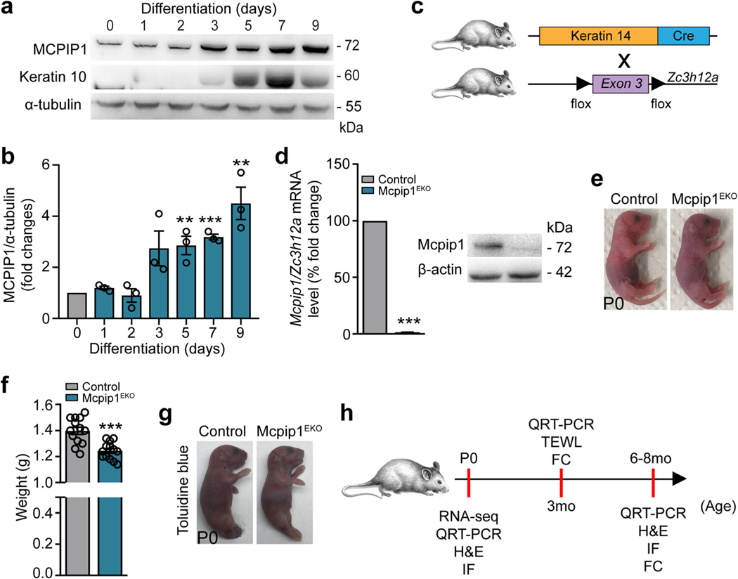
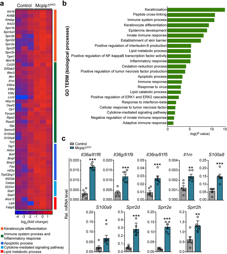
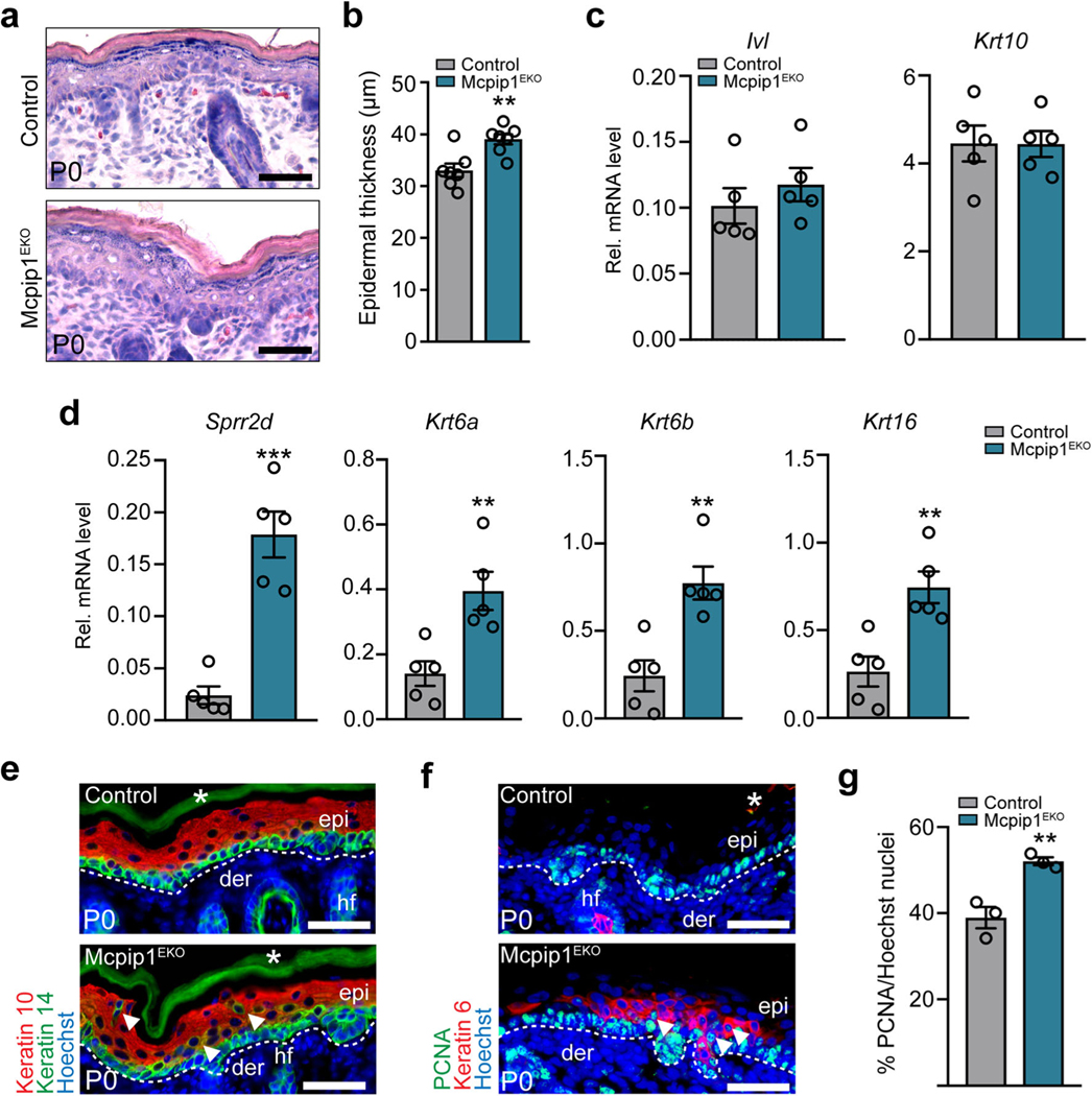
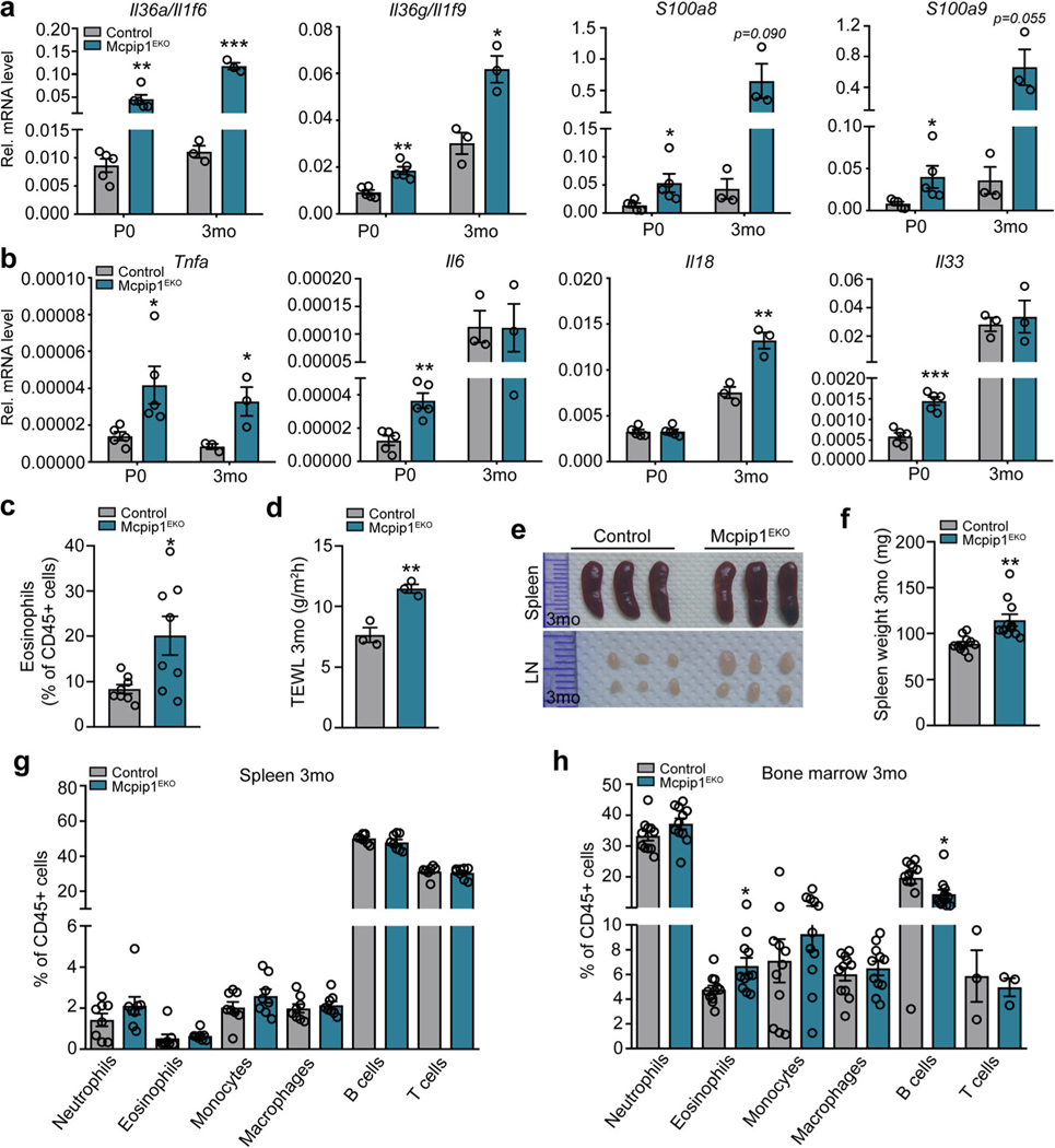
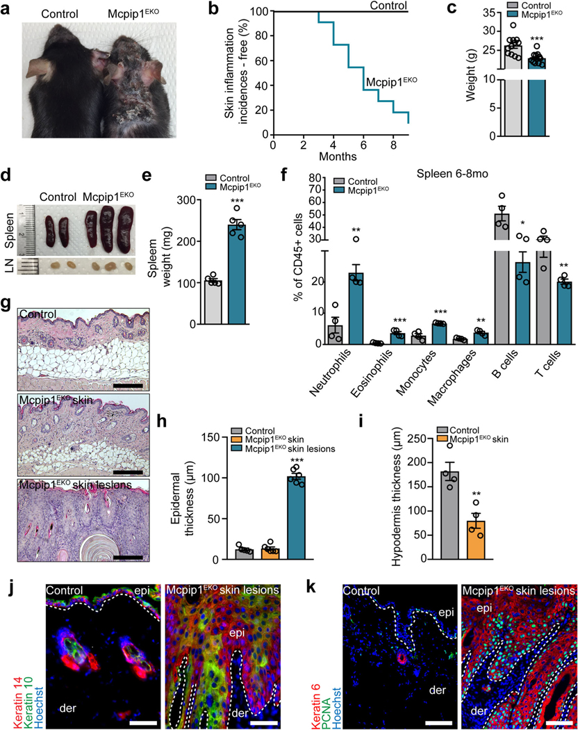
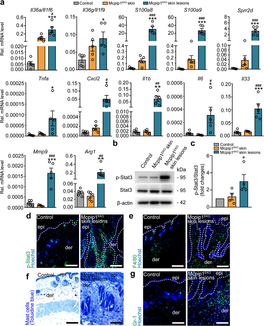
MCPIP1 (Regnase-1, encoded by the ZC3H12A gene) regulates the mRNA stability of several inflammatory cytokines. Due to the critical role of this RNA endonuclease in the suppression of inflammation, Mcpip1 deficiency in mice leads to the development of postnatal multiorgan inflammation and premature death. Here, we generated mice with conditional deletion of Mcpip1 in the epidermis (Mcpip1EKO). Mcpip1 loss in keratinocytes resulted in the upregulated expression of transcripts encoding factors related to inflammation and keratinocyte differentiation, such as IL-36α/γ cytokines, S100a8/a9 antibacterial peptides, and Sprr2d/2h proteins. Upon aging, the Mcpip1EKO mice showed impaired skin integrity that led to the progressive development of spontaneous skin pathology and systemic inflammation. Furthermore, we found that the lack of epidermal Mcpip1 expression impaired the balance of keratinocyte proliferation and differentiation. Overall, we provide evidence that keratinocyte-specific Mcpip1 activity is crucial for the maintenance of skin integrity as well as for the prevention of excessive local and systemic inflammation. KEY MESSAGES: Loss of murine epidermal Mcpip1 upregulates transcripts related to inflammation and keratinocyte differentiation. Keratinocyte Mcpip1 function is essential to maintain the integrity of skin in adult mice. Ablation of Mcpip1 in mouse epidermis leads to the development of local and systemic inflammation.

Keywords: MCPIP1; Regnase-1; Skin inflammation; ZC3H12A.


Conflict of interest statement

Conflict of interests The authors state no conflict of interests.

Figures

Fig. 1
Fig. 1
Conditional targeting of Mcpip1 in the epidermis. a Representative Western blot of three independent experiments for MCPIP1, keratin 10, and α-tubulin in primary human keratinocytes during Ca2+-induced differentiation. b Densitometric quantification of MCPIP1 levels (n = 3). c The generation of conditional Mcpip1EKO mice [26, 27]. d Left: QRT-PCR analysis of Mcpip1/Zc3h12a transcript levels (n = 7); right: Western blot for Mcpip1 and β-actin in isolated mouse keratinocytes. e Macroscopic appearance of newborn (P0) pups. f Body weights of newborn (P0) pups (n = 13). g Toluidine blue dye penetration assay at P0. h Schematic diagram representing the time points for RNA-Seq, QRT-PCR, H&E, immunofluorescence (IF), TEWL, and FC analyses. Data represent the mean ± SEM. **P < 0.01, ***P < 0.001 by unpaired t test
Fig. 2
Fig. 2
Epidermal Mcpip1 regulates the expression of genes involved in keratinocyte differentiation and the positive regulation of the immune response. RNA-Seq was performed on keratinocytes isolated from newborn Mcpip1EKO or control mice (n = 3). a Heatmap expression plot of select genes. b GO biological process terms enriched among genes with upregulated expression in Mcpip1EKO (adj. P value < 0.05 and fold change > 1.5). c QRT-PCR analysis of Il36a/Il1f6, Il36g/Il1f9, Il36ra/ Il1f5, Il1rn, S100a8, S100a9, Sprr2d, Sprr2e, and Sprr2h expression (n = 6). Data represent the mean ± SEM. *P < 0.05, **P < 0.01, ***P < 0.001 by unpaired t test
Fig. 3
Fig. 3
Newborn Mcpip1EKO mice exhibit disturbances in the expression of epidermal proliferation and differentiation markers. a H&E-stained back skin at P0. b Quantification of the epidermal thickness at P0 (n = 7). c QRT-PCR analysis of Ivl and Krt10 expression at P0 (n = 5). d QRT-PCR analysis of Sprr2d, Krt6a, Krt6b, and Krt16 expression at P0(n = 5). e Keratin 10 (Krt10) and keratin 14 (Krt14); f PCNA and keratin 6 immunofluorescence staining at P0. g Quantification of the epidermal PCNA-positive cells (n = 3). Arrowheads indicate Krt10/Krt14 double-positive suprabasal cells. Data represent the mean ± SEM. epi, epidermis; der, dermis; hf, hair follicles; *, nonspecific signal. The dashed line indicates the basal membrane. Scale bar, 100 μm. **P < 0.01, ***P < 0.001 by unpaired t test
Fig. 4
Fig. 4
Newborn and young Mcpip1EKO mice have elevated levels of inflammatory factors in their skin and develop mild systemic inflammation. QRT-PCR analysis of selected transcript levels in the control and Mcpip1EKO newborn (P0, n = 5) and young (3 mo, n = 3) mice. a Il36a/Il1f6, Il36g/Il1f9, S100a8, S100a9 transcript levels. b Tnfa, Il6, Il18, and Il33 transcript levels. c Flow cytometric analysis of 3-monthold skin eosinophils (n = 8). d Quantification of TEWL in 3-month-old mice (n = 3). e Spleenandlymph node images of 3-month-old controland Mcpip1EKO mice. f Spleen weights of 3-month-old control and Mcpip1EKO mice (n = 10). g Flow cytometric analysis of 3-month-old mouse splenic CD45+ cells (n = 8). h Flow cytometric analysis of 3-month-old mouse bone marrow CD45+ cells (n = 3–11). Data represent the mean ± SEM. *P < 0.05, **P < 0.01, ***P < 0.001 by unpaired t test
Fig. 5
Fig. 5
Development of skin lesions and systemic inflammation in old Mcpip1EKO mice. a Macroscopic appearance of 6-month-old mice. b Kaplan-Meier plots for skin inflammation incidences (n = 22). c Body weights of 6- to 8-month-old mice (n = 12). d Spleen and lymph node images of 6-month-old mice. e Spleen weights of 6- to 8-month-old mice (n = 5). f Flow cytometric analysis of 6- to 8-month-old mouse splenic CD45+ cells (n = 4). g H&E staining of control and unaffected Mcpip1EKO back skin and Mcpip1EKO skin lesions. h Quantification of the epidermal thickness (n = 6). Mcpip1EKO lesional skin was compared to control skin. i Quantification of hypodermis in 6-month-old mice (n = 4). j Keratin 14 and keratin 10; k PCNA and keratin 6 immunofluorescence staining. Data represent the mean ± SEM; epi, epidermis; der, dermis. The dashed line indicates the basal membrane. Scale bar, 100 μm. *P < 0.05, **P < 0.01, ***P < 0.001 by unpaired t test or one-way ANOVA
Fig. 6
Fig. 6
Old Mcpip1EKO mice spontaneously develop skin inflammation. a QRT-PCR analysis of Il36a/Il1f6, Il36g/Il1f9, S100a8, S100a9, Sprr2d, Tnfa, Cxcl2, Il1b, Il6, Il33, Mmp9, and Arg1 transcript levels in the healthy skin of the control and Mcpip1EKO mice (6–8 mo) and in the skin lesions of Mcpip1EKO mice (n = 5). b Representative Western blot for p-Stat3, Stat3, and β-actin in the control and Mcpip1EKO mice (6–8 mo) from four independent experiments. c Densitometric quantification of p-Stat3/Stat3 levels (n = 4). d P-Stat3 immunofluorescence staining of the skin sections. e F4/80 immunostaining. f Toluidine blue immunostaining. g Gr-1 immunostaining. Scale bar, 100 μm. Data represent the mean ± SEM; epi, epidermis; der, dermis. The dashed line indicates the basal membrane. *P < 0.05, **P < 0.01, ***P < 0.001 by one-way ANOVA. * refers to the comparison of control and Mcpip1EKO skin lesions; # refers to the comparison of Mcpip1EKO skin and Mcpip1EKO skin lesions

Similar articles

Cited by

References

    1. Nestle FO, Di Meglio P, Qin J-Z, Nickoloff BJ (2009) Skin immune sentinels in health and disease. Nat Rev Immunol 9:679–691 - PMC - PubMed
    1. Elias PM, Williams ML, Holleran WM, Jiang YJ, Schmuth M (2008) Pathogenesis of permeability barrier abnormalities in the ichthyoses: inherited disorders of lipid metabolism. J Lipid Res 49:697–714 - PMC - PubMed
    1. Guttman-Yassky E, Nograles KE, Krueger JG (2011) Contrasting pathogenesis of atopic dermatitis and psoriasis—part I: clinical and pathologic concepts. J Allergy Clin Immunol 127:1110–1118 - PubMed
    1. Segre JA (2006) Epidermal barrier formation and recovery in skin disorders. J Clin Invest 116:1150–1158 - PMC - PubMed
    1. van Smeden J, Bouwstra JA (2016) Stratum corneum lipids: their role for the skin barrier function in healthy subjects and atopic dermatitis patients. Curr Probl Dermatol 49:8–26 - PubMed

Publication types

MeSH terms

LinkOut - more resources