. 2019 Dec 1;11(1):97.
doi: 10.1186/s13195-019-0553-5.

Characterization of the selective in vitro and in vivo binding properties of crenezumab to oligomeric Aβ

Affiliations

Characterization of the selective in vitro and in vivo binding properties of crenezumab to oligomeric Aβ

William J Meilandt et al. Alzheimers Res Ther. .

Abstract

Background: Accumulation of amyloid β (Aβ) in the brain is proposed as a cause of Alzheimer's disease (AD), with Aβ oligomers hypothesized to be the primary mediators of neurotoxicity. Crenezumab is a humanized immunoglobulin G4 monoclonal antibody that has been shown to bind to synthetic monomeric and aggregated Aβ in vitro; however, less is known about the binding characteristic in vivo. In this study, we evaluated the binding patterns of crenezumab to synthetic and native forms of Aβ both in vitro and in vivo.

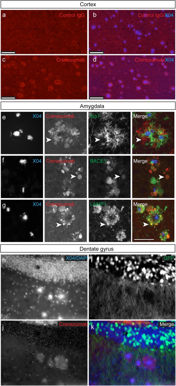
Methods: Crenezumab was used to immunoprecipitate Aβ from synthetic Aβ preparations or brain homogenates from a PS2APP mouse model of AD to determine the forms of Aβ that crenezumab interacts with. Following systemic dosing in PS2APP or nontransgenic control mice, immunohistochemistry was used to localize crenezumab and assess its relative distribution in the brain, compared with amyloid plaques and markers of neuritic dystrophies (BACE1; LAMP1). Pharmacodynamic correlations were performed to investigate the relationship between peripheral and central target engagement.

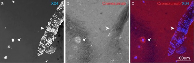
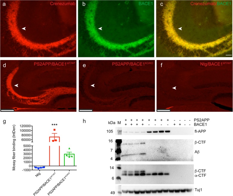
Results: In vitro, crenezumab immunoprecipitated Aβ oligomers from both synthetic Aβ preparations and endogenous brain homogenates from PS2APP mice. In vivo studies in the PS2APP mouse showed that crenezumab localizes to regions surrounding the periphery of amyloid plaques in addition to the hippocampal mossy fibers. These regions around the plaques are reported to be enriched in oligomeric Aβ, actively incorporate soluble Aβ, and contribute to Aβ-induced neurotoxicity and axonal dystrophy. In addition, crenezumab did not appear to bind to the dense core region of plaques or vascular amyloid.

Conclusions: Crenezumab binds to multiple forms of amyloid β (Aβ), particularly oligomeric forms, and localizes to brain areas rich in Aβ oligomers, including the halo around plaques and hippocampal mossy fibers, but not to vascular Aβ. These insights highlight a unique mechanism of action for crenezumab of engaging Aβ oligomers.

Keywords: Alzheimer’s disease; Amyloid β; Crenezumab; Mossy fiber; Oligomeric; Vascular amyloid.


Conflict of interest statement

All authors are full-time employees of Genentech, Inc., and shareholders in F. Hoffmann-La Roche Ltd. The authors declare that they have no competing interests.

Figures

Fig. 1
Fig. 1
Crenezumab recognizes Aβ oligomers from in vitro and in vivo sources. Pre-formed (m)onomericAβ42, (o)ligomericAβ42, or (a)ggregatedAβ42 were run on native PAGE at 1000, 500, and 250 ng per lane to visualize Aβ42 banding patterns (a). Note that aAβ was too large to enter the gel. Antibodies were incubated with pre-formed Aβ42 oligomers overnight at 4 °C. To visualize nondenatured oligomers, immunoprecipitated (IP) eluates using were run on native PAGE. Crenezumab recognizes both low molecular weight oligomers between 20 and 50 kDa and high molecular weight (HMW) oligomers between 250 and 700 kDa (b). Anti-Aβ IPs from the soluble fraction of PS2APP mouse brain homogenates were run on native PAGE. Crenezumab recognizes HMW oligomers (c). 6E10 and 4G8 were used as detection antibodies on all blots
Fig. 2
Fig. 2
In vivo-dosed crenezumab binds in a halo around amyloid plaques and to dystrophic neurites in PS2APP mice. In vivo-dosed crenezumab (200 mg/kg, i.v.) was visualized 7 days postdose with anti-hIgG-Alexa594 antibody (red), and plaques were stained with methoxy-X04 (blue). Representative epifluorescent images of plaque-associated halo of staining by crenezumab alone (c) and with plaques (d) in the cortex. Note the absence of staining in the control-injected (control IgG, gD) mice around plaques (a, b). In the amygdala (eg), confocal z-stacked images show crenezumab binding was prominent around the core of the plaque but in regions not covered by microglia (e) (labeled with Iba1, green). This staining pattern was reminiscent of dystrophic neurites and was confirmed by co-staining of crenezumab (80 mg/kg, i.v., red) with markers of dystrophic neurites including BACE1 (green, f) and LAMP1 (green, g). Arrowheads indicate example regions of overlap. In vivo-dosed crenezumab (j, k, red, 120 mg/kg, IP) was localized to regions between methoxy-X04-labeled plaques (h, k, blue) and GFP-labeled dendrites (i, k, green) in the dentate gyrus of PS2APP-GFP (line M) mice (2 days postdose). Scale bar, 200 μm (ad) and 50 μm (eg)
Fig. 3
Fig. 3
In vivo-dosed crenezumab does not bind to vascular amyloid in PS2APP mice. Representative confocal × 40 images (z-stack maximum projection) of parenchymal amyloid plaques (arrow) and vascular amyloid (arrowhead) stained with methoxy-X04 (a, c, blue). Note the selective staining of in vivo-dosed crenezumab (200 mg/kg, i.v.) (b, c, red) to the peri-plaque region and the absence from the vascular amyloid. Scale bar, 100 μm
Fig. 4
Fig. 4
In vivo-dosed crenezumab binds to the mossy fibers in PS2APP mice. In vivo-dosed crenezumab, but not control IgG (anti-gD IgG4), dose-dependently binds to the mossy fiber axons in the hippocampus of PS2APP mice. Representative epifluorescent images of mossy fiber binding by crenezumab in PS2APP mice (a). Quantification of mossy fiber binding integrated density (IntDen) found a significant treatment effect (b) (ANOVA: F4,19 = 50.10, p < 0.0001). ANOVA followed by Tukey’s multiple comparison test: *p < 0.05, ***p < 0.001, ****p < 0.0001 as indicated or to control IgG (anti-gD)
Fig. 5
Fig. 5
Crenezumab binding to the hippocampal mossy fibers is Aβ dependent. Representative epifluorescent images of in vivo-dosed crenezumab (80 mg/kg) binding to the mossy fibers (a) of PS2APP mice (arrows). Immunostaining for BACE1 shows strong binding in the mossy fibers (b) that overlap with crenezumab staining (c, merge). Scale bar = 50 μm. In vivo-dosed crenezumab (80 mg/kg) staining to the mossy fibers in the PS2APP/BACE1WT/WT mice (d) was nearly completely absent in PS2APP/BACE1KO/KO (e) compared with Ntg/BACE1WT/WT (f) mice. Scale bar, 200 μm. g Significant differences in mossy fiber binding were found between the groups (ANOVA: F2,8 = 29.16, p < 0.001) n = 3–4/group. ANOVA followed by Tukey’s multiple comparison test. ***p < 0.001 versus all others. h Western blots of full-length/soluble APP and Aβ (detected by 4G8 and 6E10) and α/β–C-terminal fragment (detected by SIG-39152) from soluble hippocampal TBS homogenates from PS2APP/BACE1WT/WT, PS2APP/BACE1KO/KO, and Ntg/BACE1WT/WT mice. β-Tubulin (Tuj1) was used as a loading control. M, molecular weight marker
Fig. 6
Fig. 6
In vivo-dosed crenezumab binds to (o)ligomeric Aβ, not to (mo)nomeric Aβ, in the hippocampal mossy fiber tract. Plasma and cerebellum PK levels 6 h after the final day of dosing (100 mg/kg daily for 5 d) with an anti-moAβ (n = 3) or 5 days after a single injection of control IgG (anti-gD 40 mg/kg, n = 4) or crenezumab (80 mg/kg, n = 4) in PS2APP mice. ANOVA found a significant difference in plasma PK levels (a) (F2,8 = 86.90, p < 0.0001) but not in the cerebellum (b; not significant [NS]). Quantification (c) and representative epifluorescent images (df) of mossy fiber binding by crenezumab but not by control IgG or moAβ antibodies. ANOVA found a significant difference in binding (F2,8 = 26.84, p < 0.001). Representative images of ex vivo oAβ staining in the mossy fibers (arrows) of PS2APP mice (g), but not in Ntg mice (h), using an anti-oAβ antibody (mab-M). Scale bar, 50 μm. ANOVA followed by Tukey’s multiple comparison test: *p < 0.05, ***p < 0.001, ****p < 0.0001 as indicated or to control IgG
Fig. 7
Fig. 7
In vivo crenezumab-mediated target engagement correlates between the periphery and brain. Significant elevations in plasma Aβ40 (a) (ANOVA: F4,19 = 23.43, p < 0.0001) and Aβ42 (b) (ANOVA: F4,19 = 12.08, p < 0.0001) were found 7 days after crenezumab treatment. A significant correlation was observed between the elevations in plasma Aβ40 (c) (R2 = 0.08, p < 0.001) and Aβ42 (d) (R2 = 0.67, p < 0.001) and crenezumab mossy fiber binding (see Fig. 4) by linear regression. N = 5/group. ANOVA followed by Tukey’s multiple comparison test: *p < 0.05, **p < 0.01, ***p < 0.001, ****p < 0.0001 as indicated or to control IgG

References

    1. Selkoe DJ, Hardy J. The amyloid hypothesis of Alzheimer’s disease at 25 years. EMBO Mol Med. 2016;8:595–608. - PMC - PubMed
    1. Haass C, Selkoe DJ. Soluble protein oligomers in neurodegeneration: lessons from the Alzheimer’s amyloid beta-peptide. Nat Rev Mol Cell Biol. 2007;8:101–112. - PubMed
    1. Benilova I, Karran E, De Strooper B. The toxic Aβ oligomer and Alzheimer’s disease: an emperor in need of clothes. Nat Neurosci. 2012;15:349–357. - PubMed
    1. Mucke L, Selkoe D. Neurotoxicity of amyloid β-protein: synaptic and network dysfunction. Cold Spring Harb Perspect Med. 2012;2:a006338. - PMC - PubMed
    1. Lesne S, Koh MT, Kotilinek L, Kayed R, Glabe CG, Yang A, et al. A specific amyloid-beta protein assembly in the brain impairs memory. Nature. 2006;440:352–357. - PubMed

Publication types