Review
. 2020 Jan;43(1):42-54.
doi: 10.1016/j.tins.2019.11.002. Epub 2019 Nov 29.

Neocortex-Cerebellum Circuits for Cognitive Processing

Affiliations
Review

Neocortex-Cerebellum Circuits for Cognitive Processing

Mark J Wagner et al. Trends Neurosci. 2020 Jan.

Abstract

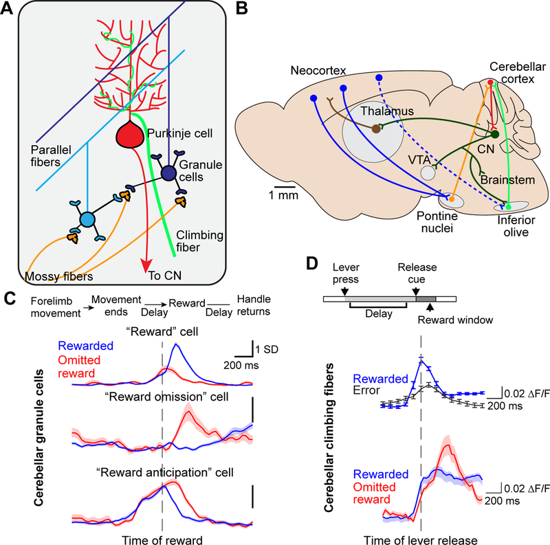
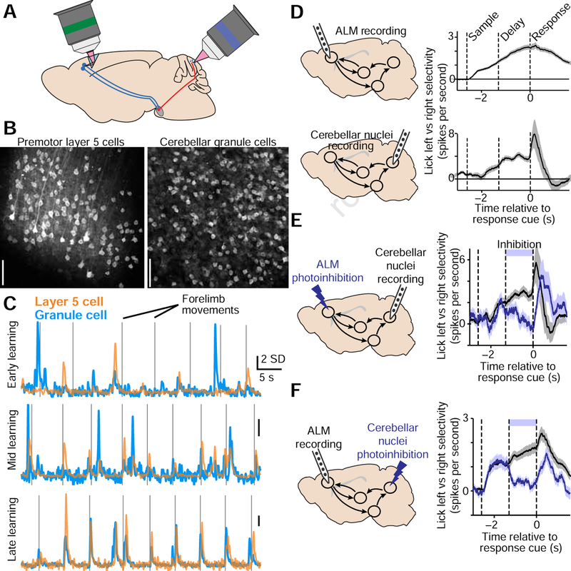
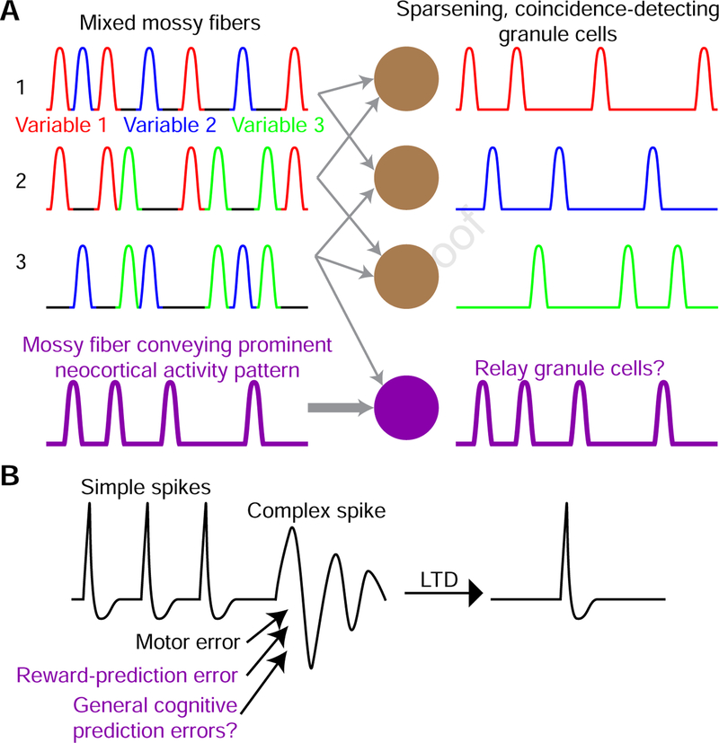
Although classically thought of as a motor circuit, the cerebellum is now understood to contribute to a wide variety of cognitive functions through its dense interconnections with the neocortex, the center of brain cognition. Recent investigations have shed light on the nature of cerebellar cognitive processing and information exchange with the neocortex. We review findings that demonstrate widespread reward-related cognitive input to the cerebellum, as well as new studies that have characterized the codependence of processing in the neocortex and cerebellum. Together, these data support a view of the neocortex-cerebellum circuit as a joint dynamic system both in classical sensorimotor contexts and reward-related, cognitive processing. These studies have also expanded classical theory on the computations performed by the cerebellar circuit.

Keywords: cerebellum; learning; motor control; neocortex; neural dynamics; reward.


Figures

Figure 1.
Figure 1.. Cerebellar circuits and reward signals
A. Cerebellar cortex microcircuit. Input arrives via the mossy fiber pathway from specific nuclei in the pons, medulla, and spinal cord, as well as from the climbing fiber pathway from the inferior olive. Each mossy fiber synapses onto ~50 granule cells, and each granule cell receives input from about four mossy fibers. ~100,000 granule cell parallel fiber axons synapse onto each Purkinje cell, while each Purkinje cell receives input from only one climbing fiber. For simplicity, GABAergic interneurons in the cerebellar cortex are omitted from this schematic. Purkinje cells project axons to the cerebellar nuclei (CN), which also receive collaterals from both mossy fibers and climbing fibers (not shown). B. Connections between the cerebellum and other brain regions. Nearly all subcortically-projecting layer 5 pyramidal neurons throughout the neocortex send an axon collateral to the pontine nuclei. The cerebellum receives mossy fibers from all pontine nuclei neurons, and climbing fibers from inferior olive neurons. The cerebellar nuclei (CN), targets of Purkinje cell axons, project to numerous targets including the cortex via the thalamus, the ventral tegmental area (VTA), and the brainstem nuclei including the inferior olive. Dashed arrows represent indirect input from the cortex to the inferior olive. C. Reward expectation signaling in cerebellar granule cells. Top, mice executed a forelimb operant task for water reward during two-photon Ca2+ imaging [38]. Bottom, three example granule cell activity profiles. Traces show fluorescence aligned to reward delivery (dashed vertical line) and averaged across either rewarded trials or trials on which reward was unexpectedly omitted. From top to bottom, these three cells were active preferentially during reward delivery, reward omission, or the delay while the mouse waited for the reward. Note that the “reward anticipation” cell remained active longer while the mouse continued waiting following unexpected reward omission, until the mouse gave up and ceased licking (not shown). D. Reward expectation signaling in cerebellar climbing fibers. Top, water-restricted mice executed a press-and-hold forelimb lever task for water reward during two-photon Ca2+ imaging of Purkinje dendrites [52], whose fluorescence reports climbing fiber activity, in lobule simplex of the cerebellum. Bottom, example climbing fiber activity. Climbing fibers became active preferentially at the time of the lever release on correctly-timed trials, which predicted subsequent reward, but not on error trials, which did not yield subsequent reward. On correctly-timed trials on which the subsequent reward was omitted, a second climbing fiber response was elicited. C reproduced with permission from [38], D reproduced with permission from [52].
Figure 2.
Figure 2.. Neocortex-cerebellum communication
A. Schematic of strategy for simultaneous two-photon Ca2+ imaging of both premotor cortical layer 5 pyramidal neurons of the rostral forelimb area, and cerebellar granule cells, during a forelimb movement planning task. B. Mean two-photon brain images of the premotor cortex and cerebellum showing detected layer 5 cells and granule cells highlighted in grayscale. C. An example layer 5–granule cell pair that was tracked over several weeks of task learning. Traces show time-varying fluorescence magnitude. Over learning, this pair develops strongly correlated activity. SD, standard deviation used as unit for these z-scored traces. D. Mice discriminated two whisker stimuli and reported stimulus identity by licking left or right after an enforced delay period. Top, recordings from the anterior lateral motor area (ALM) during the task. ALM neurons develop stimulus selectivity during the sample period (between the left and middle dashed lines) that persists through the delay period (between the middle and right dashed lines) and into the response period. Bottom, recordings from the cerebellar output nuclei reveal a similar timeline of stimulus selectivity. E,F. Cerebellar nuclei recordings during ALM photoinhibition (E) and ALM recordings during cerebellar nuclei photoinhibition (F) demonstrated that directional selectivity in either brain region requires intact signaling in the other region. A-C reproduced with permission from [72]. D,E, reproduced with permission from [75].
Figure 3.
Figure 3.. Computational theories of cerebellar function.
A. Top, in classical theory, granule cells threshold their approximately four inputs (here, will spike only if two inputs are simultaneously active), in order to perform coincidence detection. Since the number of granule cells far exceeds the number of unique mossy fiber inputs, if each granule cell integrates a unique combination of four randomly chosen mossy fibers, the resulting granule cell ensemble output will be sparse and high dimensional. This allows Purkinje cells to discriminate between activity that appears very similar at the level of the mixed mossy fibers. Bottom (purple), during motivated behavior, some granule cells appear to be densely active in ways that faithfully recapitulate the selectivity and tuning of individual cortical signals relayed by a putative task-critical mossy fiber. B. Left, a Purkinje cell’s simple spikes are periodically interspersed with complex spikes from the climbing fiber input. In classical theory, complex spikes are triggered by motor errors, which indicate that the previous simple spiking was inappropriate, and thus produces long term depression (LTD) at the Purkinje cell’s synaptic inputs from the set of parallel fibers that were active in the previous several hundred milliseconds (right). New evidence indicates that events that predict upcoming reward, in addition to a violation of that prediction via a reward omission, both can elicit complex spiking. This may be consistent with an unsigned reward prediction error signal, while opening the door to a potential range of other types of cognitive prediction error signals.

References

    1. Ito M (2000) Mechanisms of motor learning in the cerebellum. Brain research 886, 237–245 - PubMed
    1. Ivry RB, et al. (2002) The cerebellum and event timing. Ann NY Acad Sci 978, 302–317 - PubMed
    1. Bastian AJ (2006) Learning to predict the future: the cerebellum adapts feedforward movement control. Current Opinion in Neurobiology 16, 645–649 - PubMed
    1. Gao Z, et al. (2012) Distributed synergistic plasticity and cerebellar learning. Nat Rev Neurosci 13, 619. - PubMed
    1. Diedrichsen J, et al. (2019) Universal Transform or Multiple Functionality? Understanding the Contribution of the Human Cerebellum across Task Domains. Neuron 102, 918–928 - PMC - PubMed

Publication types