Review
. 2020 Mar;45(3):244-258.
doi: 10.1016/j.tibs.2019.11.001. Epub 2019 Nov 29.

The SLC25 Mitochondrial Carrier Family: Structure and Mechanism

Affiliations
Review

The SLC25 Mitochondrial Carrier Family: Structure and Mechanism

Jonathan J Ruprecht et al. Trends Biochem Sci. 2020 Mar.

Abstract

Members of the mitochondrial carrier family (SLC25) provide the transport steps for amino acids, carboxylic acids, fatty acids, cofactors, inorganic ions, and nucleotides across the mitochondrial inner membrane and are crucial for many cellular processes. Here, we use new insights into the transport mechanism of the mitochondrial ADP/ATP carrier to examine the structure and function of other mitochondrial carriers. They all have a single substrate-binding site and two gates, which are present on either side of the membrane and involve salt-bridge networks. Transport is likely to occur by a common mechanism, in which the coordinated movement of six structural elements leads to the alternating opening and closing of the matrix or cytoplasmic side of the carriers.

Keywords: adenine nucleotide translocase; energetics; mitochondrial transport; salt-bridge networks; transport mechanism; uncoupling protein.


Figures

Figure 1
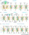
Figure 1. Selected Transporters of the Human Mitochondrial Carrier Family (SLC25).
Mitochondrial carriers transport (A) amino acids; (B), nucleotides, cofactors, and inorganic ions; and (C) protons, fatty acids, and di- and tricarboxylates across the mitochondrial inner membrane using antiport, symport, and uniport activities [78]. The ATP-Mg/Pi carrier and aspartate glutamate carriers have a three-domain structure, comprising a calcium-regulated domain, a carrier domain, and a domain with an amphipathic helix. Protons are shown as grey spheres with a plus sign. The structures are homology models based upon PDB: 1OKC and 4C9H chain A, and generated by Swiss-Model [79]. The regulatory domains of AGC and APC are based upon PDB: 4P5W and 4ZCU, respectively, and the links to the carrier domains are not modelled. Abbreviations: AAC, ADP/ATP carrier (SLC25A4, SLC25A5, SLC25A6, SLC25A31); AGC, aspartate glutamate carrier (SLC25A12, SLC25A13); APC, ATP-Mg/Pi carrier (SLC25A23, SLC25A24, SLC25A25); BAC, basic amino acid carrier (SLC25A29); CAC, carnitine-acylcarnitine carrier (SLC25A20); CIC, citrate (tricarboxylate) carrier (SLC25A1); DIC, dicarboxylate carrier (SLC25A10); GC, glutamate carrier (SLC25A18, SLC25A22); GLYC: glycine carrier (SLC25A38); IM inner membrane; IS, intermembrane space; MM mitochondrial matrix; MTFRN mitoferrin (SLC25A28, SLC25A37); ODC, oxoadipate carrier (SLC25A21); OGC, oxoglutarate carrier (SLC25A11); ORC, ornithine carrier (SLC25A2, SLC25A15); PIC, phosphate carrier (SLC25A3); SAMC, S-adenosylmethionine carrier (SLC25A26); TPC, thiamine pyrophosphate carrier (SLC25A19); UCP1, uncoupling protein (SLC25A7).
Figure 2
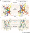
Figure 2. Key Structural and Functional Elements of Mitochondrial Carriers.
Structures of (A) the cytoplasmic state (c-state) of the mitochondrial ADP/ATP carrier trapped by the inhibitor carboxyatractyloside (CATR) (ScAac2p, PDB: 4C9H chain A [49]); and (B) the matrix state (m-state) ADP/ATP carrier trapped by the inhibitor bongkrekic acid (BKA) (TtAac, PDB: 6GCI chain A [54]). The proteins are shown in cartoon representation (blue, domain 1; yellow, domain 2; red, domain 3). Transmembrane α-helices (H1–H6) and matrix α-helices (h12, h34, and h56) are indicated. The inhibitors are shown as space-filling models, with blue-green carbon atoms for CATR and orange carbon atoms for BKA. The cardiolipin molecules are shown in ball-and-stick representation with dark grey, red, and orange balls for carbon, oxygen, and phosphorus atoms, respectively. Key functional elements, highlighted in different colors, in the (C) c- and (D) m-states, viewed in the same orientation as (A) and (B), respectively.
Figure 3
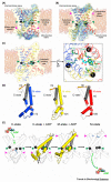
Figure 3. Substrate-Binding and Conformational Changes between Cytoplasmic- and Matrix-States.
(A) The c-state of the mitochondrial ADP/ATP carrier (ScAac2p, PDB: 4C9H chain A), showing the substrate-binding site residues (green sticks) and matrix gate, and with the water-accessible surface in pale blue. (B) The m-state of the mitochondrial ADP/ATP carrier (TtAac, PDB: 6GCI chain A), showing the substrate-binding site residues (green sticks) and cytoplasmic gate, with the water-accessible surface in pale blue. (C) ATP docked into the proposed substrate-binding site of the m-state. Interactions between ATP and substrate-binding site residues are indicated by yellow dashed lines in the right-hand panel. (D) Comparison of the domain structures of the c-state, shown in outline, and m-state, shown with domains 1–3 colored blue, yellow, and red, respectively. The prolines of the conserved Px[DE]xx[KR] signature motifs are indicated by brown spheres. Orange arrows indicate the inward movement of the gate elements (shown in grey). (E) Conformational changes between c- and m-states, viewed laterally from the membrane. Matrix and cytoplasmic network residues are shown as sticks in the left- and right-hand panels, respectively. Conformational changes induced by substrate-binding involve (1) rotations of the core elements of each domain (highlighted here for domain 2), shown by a black arrow, and (2) inward movements of the gate elements (grey arrow). In (A–C) and (E), the position of the substrate-binding site is highlighted by green arrowheads and the gates are highlighted by magenta arrowheads. In (A–E), the contact points of the substrate-binding site are indicated by black spheres with Roman numerals.
Figure 4
Figure 4. Relative Cytoplasmic and Matrix Network Strengths of Different SLC25 Family Members.
Positively charged, negatively charged, polar, aliphatic, and aromatic residues are shown in blue, red, green, pink, and cyan colors, respectively. The bonds are shown as black dashes. The number in the center is the total interaction energy of the network, where salt-bridge interactions and braces are counted as 1.0 and 0.5, respectively. The models of the cytoplasmic network are derived from PDB:6GCI (chain A), whereas those of the matrix network are derived from PDB:1OKC.
Figure 5
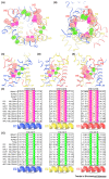
Figure 5. Conserved πGπxπG and πxxxπ Motifs Allow Close-Packing of Helices in the Matrix State (m-State).
(A) Structure of the cytoplasmic state (c-state) of the mitochondrial ADP/ATP carrier (ScAac2p, PDB: 4C9H chain A); and (B) structure of the m-state (TtAac, PDB: 6GCI chain A), both viewed from the cytoplasmic side of the membrane (blue, domain 1; yellow, domain 2; red, domain 3). The motifs are shown in stick representation and with semitransparent spheres at the van der Waals radius of the appropriate atom. The πGπxπG motif is colored salmon and magenta at the positions of π and G residues, respectively. The πxxxπ motif is colored green at the position of the π residues. (C-E) Side views of the motifs at domain interfaces in the m-state, highlighting domain 1 (C), domain 2 (D), and domain 3 (E). (F) Amino acid sequences of selected mitochondrial carriers around the πGπxπG motif on H1 (left), H3 (middle), and H5 (right). (G) Amino acid sequences of selected mitochondrial carriers around the πxxxπ motif on H2 (left), H4 (middle), and H6 (right). In (F) and (G), residues of the motifs are in boxes colored as in (A) to (E). Abbreviations: AAC, ADP/ATP carrier; AGC, aspartate glutamate carrier; APC, ATP-Mg/Pi carrier; CAC, carnitine-acylcarnitine carrier; CIC, citrate (tricarboxylate) carrier; OGC, oxoglutarate carrier; PIC, phosphate carrier; TPC, thiamine pyrophosphate carrier; UCP1, uncoupling protein.
Figure 6
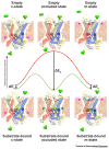
Figure 6. Structures and Energetics of Intermediates in the Mitochondrial Carrier Transport Cycle.
The structures of the cytoplasmic (c-), matrix (m-) and occluded states are based upon models of the uninhibited mitochondrial ADP/ATP carrier [54] and are shown with cylindrical helices, colored blue, yellow, and red for the core elements of domains 1, 2, and 3, respectively, with the gate elements in grey. The cytoplasmic and matrix gate residues are shown as sticks, with interactions shows as magenta dots when the gates are closed (magenta arrowheads). The contact points of the substrate-binding site (green arrowheads) are shown as black spheres with Roman numerals. Substrate (green trigonal object) binding to the carrier reduces the energy state of the intermediates. This is most pronounced for the occluded state, the substrate-binding site of which optimally interact with the substrate, lowering the energy barrier for transport. Figure adapted, with permission, from [71].

References

    1. Palmieri F, Klingenberg M. On the possible role of structural protein in the binding and translocation of adenine nucleotides in mitochondria. Biochim Biophys Acta. 1967;131:582–585. - PubMed
    1. Aquila H, et al. Complete amino acid sequence of the ADP/ATP carrier from beef heart mitochondria. Hoppe Seylers Z Physiol Chem. 1982;363:345–349. - PubMed
    1. Klingenberg M. The ADP and ATP transport in mitochondria and its carrier. Biochim Biophys Acta. 2008;1778:1978–2021. - PubMed
    1. Kunji ERS, et al. The transport mechanism of the mitochondrial ADP/ATP carrier. Biochim Biophys Acta. 2016;1863:2379–2393. - PubMed
    1. Ruprecht JJ, Kunji ERS. Structural changes in the transport cycle of the mitochondrial ADP/ATP carrier. Curr Opin Struct Biol. 2019;57:135–144. - PMC - PubMed

Publication types

Substances

LinkOut - more resources