

Seeding of a Tumor in the Gastric Wall after Endoscopic Ultrasound-guided Fine-needle Aspiration of Solid Pseudopapillary Neoplasm of the Pancreas
- PMID: 31787691
- PMCID: PMC7118382
- DOI: 10.2169/internalmedicine.3244-19
Seeding of a Tumor in the Gastric Wall after Endoscopic Ultrasound-guided Fine-needle Aspiration of Solid Pseudopapillary Neoplasm of the Pancreas
Abstract
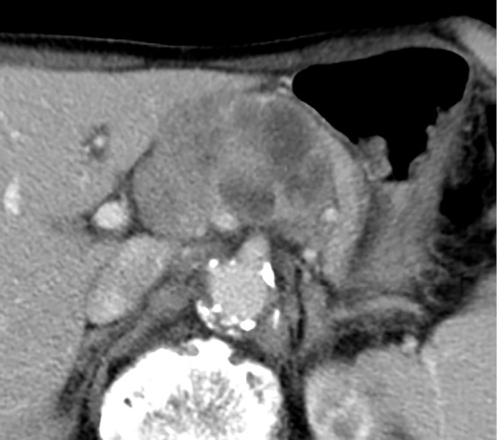
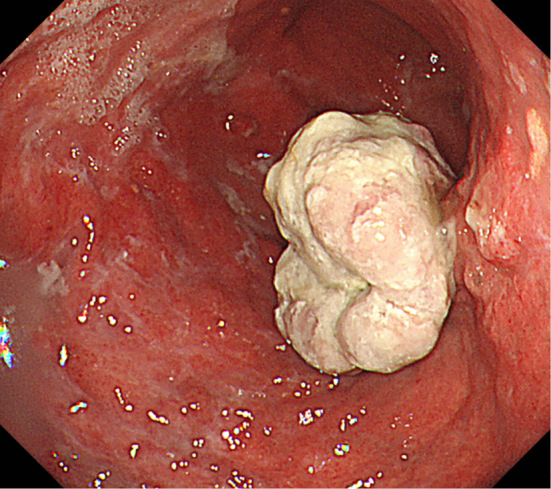
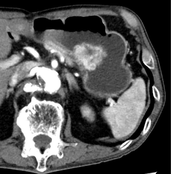
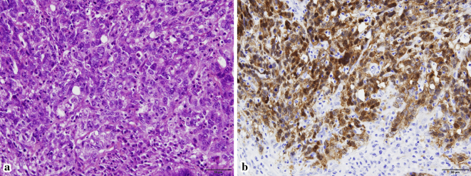
Endoscopic ultrasound-guided fine-needle aspiration (EUS-FNA) is widely used as a first-line procedure for the definitive diagnosis of pancreatic solid tumor. Adverse events associated with the EUS-FNA procedure include acute pancreatitis, bleeding, infection, and duodenal perforation. Rarely, pancreatic tumors disseminate in the peritoneal cavity or seed in the gastric wall via the biopsy needle tract after EUS-FNA. Such seeding has been noted primarily in cases of adenocarcinomas and has not been associated with solid pseudopapillary neoplasm (SPN), a rare and potentially malignant tumor of the pancreas. This is the first report of a case of tumor seeding in the gastric wall after EUS-FNA of pancreatic SPN.
Keywords: endoscopic ultrasound-guided fine-needle aspiration; needle tract seeding; solid pseudopapillary neoplasm.
Conflict of interest statement
Figures

References
-
- Dumonceau JM, Polkowski M, Larghi A, et al. . Indications, results, and clinical impact of endoscopic ultrasound (EUS)-guided sampling in gastroenterology: European Society of Gastrointestinal Endoscopy (ESGE) Clinical Guideline. Endoscopy 43: 897-912, 2011. - PubMed
-
- Wang KX, Ben QW, Jin ZD, et al. . Assessment of morbidity and mortality associated with EUS-guided FNA: a systematic review. Gastrointest Endosc 73: 283-290, 2011. - PubMed
-
- Yoon WJ, Daglilar ES, Fernandez-del Castillo C, Mino-Kenudson M, Pitman MB, Brugge WR. Peritoneal seeding in intraductal papillary mucinous neoplasm of the pancreas patients who underwent endoscopic ultrasound-guided fine-needle aspiration: the PIPE Study. Endoscopy 46: 382-387, 2014. - PubMed