

Mechanical stimulation of growth plate chondrocytes: Previous approaches and future directions
- PMID: 31787777
- PMCID: PMC6884322
- DOI: 10.1007/s11340-018-0424-1
Mechanical stimulation of growth plate chondrocytes: Previous approaches and future directions
Abstract
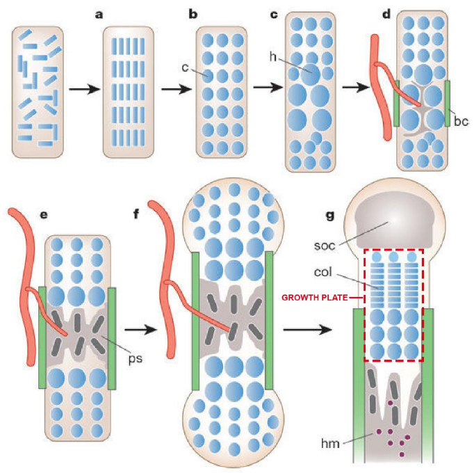
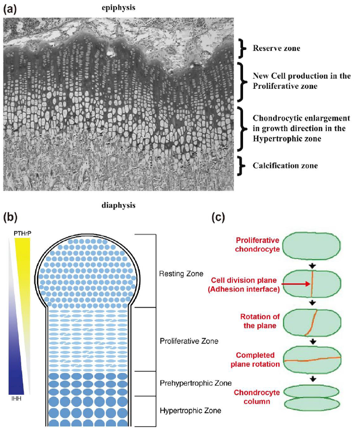
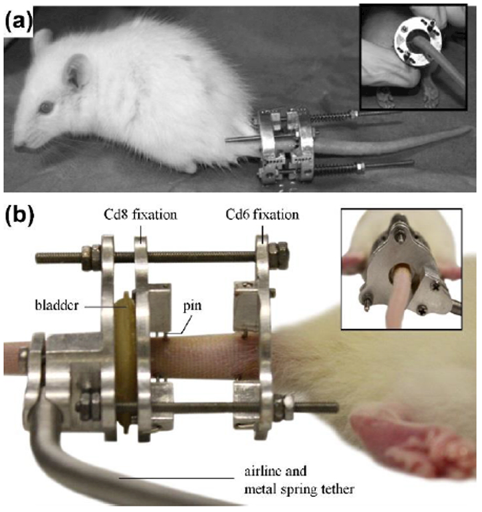
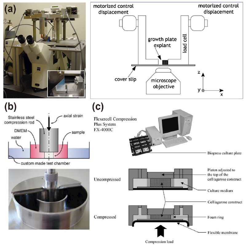
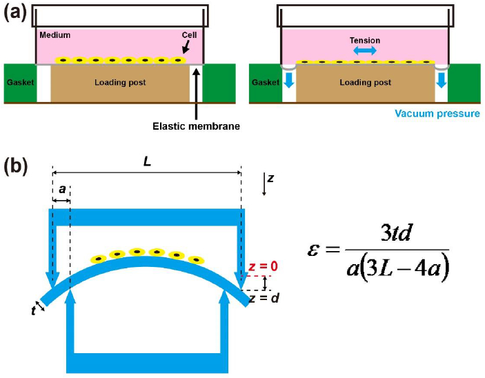
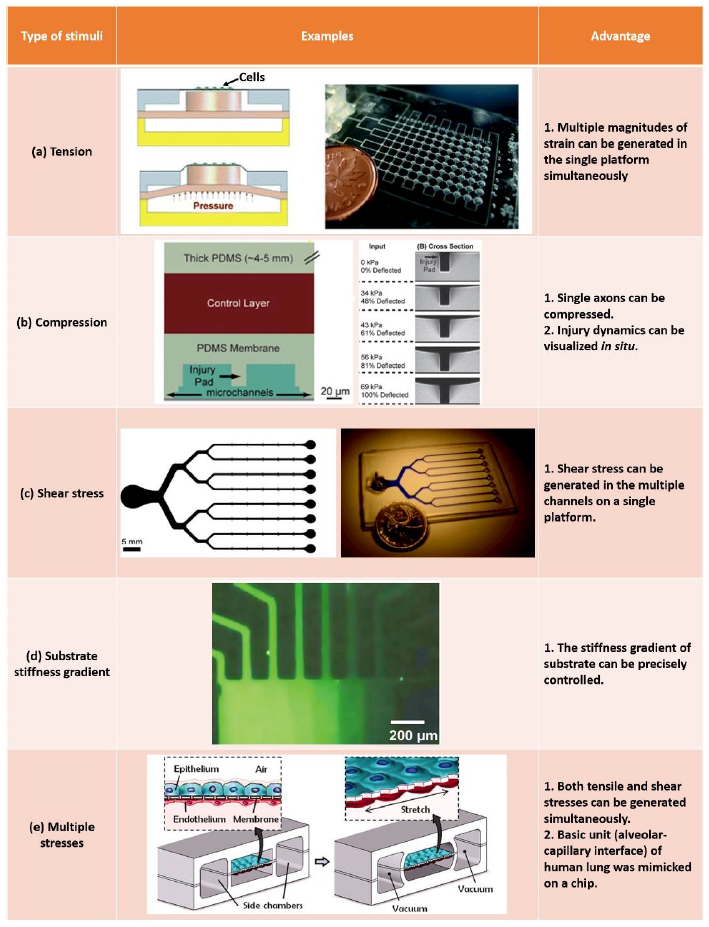
Growth plate cartilage resides near the ends of long bones and is the primary driver of skeletal growth. During growth, both intrinsically and extrinsically generated mechanical stresses act on chondrocytes in the growth plate. Although the role of mechanical stresses in promoting tissue growth and homeostasis has been strongly demonstrated in articular cartilage of the major skeletal joints, effects of stresses on growth plate cartilage and bone growth are not as well established. Here, we review the literature on mechanobiology in growth plate cartilage at macroscopic and microscopic scales, with particular emphasis on comparison of results obtained using different methodological approaches, as well as from whole animal and in vitro experiments. To answer these questions, macroscopic mechanical stimulators have been developed and applied to study mechanobiology of growth plate cartilage and chondrocytes. However, the previous approaches have tested a limited number of stress conditions, and the mechanobiology of a single chondrocyte has not been well studied due to limitations of the macroscopic mechanical stimulators. We explore how microfluidics devices can overcome these limitations and improve current understanding of growth plate chondrocyte mechanobiology. In particular, microfluidic devices can generate multiple stress conditions in a single platform and enable real-time monitoring of metabolism and cellular behavior using optical microscopy. Systematic characterization of the chondrocytes using microfluidics will enhance our understanding of how to use mechanical stresses to control the bone growth and the properties of tissue-engineered growth plate cartilage.
Keywords: Growth plate chondrocyte; bone growth; mechanobiology; microfluidics.
Conflict of interest statement
The authors declare no competing financial interest.
Figures
Similar articles
-
Pneumatic microfluidic cell compression device for high-throughput study of chondrocyte mechanobiology.Lab Chip. 2018 Jul 10;18(14):2077-2086. doi: 10.1039/c8lc00320c. Lab Chip. 2018. PMID: 29897088 Free PMC article.
-
Effects of shear stress on articular chondrocyte metabolism.Biorheology. 2000;37(1-2):95-107. Biorheology. 2000. PMID: 10912182 Review.
-
A Microfluidic Platform for Stimulating Chondrocytes with Dynamic Compression.J Vis Exp. 2019 Sep 13;(151):10.3791/59676. doi: 10.3791/59676. J Vis Exp. 2019. PMID: 31566611 Free PMC article.
-
Importance of collagen orientation and depth-dependent fixed charge densities of cartilage on mechanical behavior of chondrocytes.J Biomech Eng. 2008 Apr;130(2):021003. doi: 10.1115/1.2898725. J Biomech Eng. 2008. PMID: 18412490
-
Biomechanical properties and mechanobiology of the articular chondrocyte.Am J Physiol Cell Physiol. 2013 Dec 15;305(12):C1202-8. doi: 10.1152/ajpcell.00242.2013. Epub 2013 Sep 25. Am J Physiol Cell Physiol. 2013. PMID: 24067919 Review.
Cited by
-
Mechanical stimulation in 2D: A potent accelerator of matrix mineralization in ATDC5 chondrogenic cells.J Orthop. 2025 May 31;70:173-182. doi: 10.1016/j.jor.2025.05.058. eCollection 2025 Dec. J Orthop. 2025. PMID: 40546983
-
Development of the mechanoresponsive pericellular matrix of chondrons.Sci Adv. 2025 May 2;11(18):eado6644. doi: 10.1126/sciadv.ado6644. Epub 2025 May 2. Sci Adv. 2025. PMID: 40315311 Free PMC article.
-
Mucopolysaccharidosis IVA: Current Disease Models and Drawbacks.Int J Mol Sci. 2023 Nov 9;24(22):16148. doi: 10.3390/ijms242216148. Int J Mol Sci. 2023. PMID: 38003337 Free PMC article. Review.
-
Growing Pains: The Need for Engineered Platforms to Study Growth Plate Biology.Adv Healthc Mater. 2022 Oct;11(19):e2200471. doi: 10.1002/adhm.202200471. Epub 2022 Aug 15. Adv Healthc Mater. 2022. PMID: 35905390 Free PMC article. Review.
-
Novel dual: rod plate system for EOS improves vertebral wedging and permits spinal growth.J Orthop Surg Res. 2023 Sep 29;18(1):738. doi: 10.1186/s13018-023-04094-9. J Orthop Surg Res. 2023. PMID: 37773144 Free PMC article.
References
-
- Kronenberg HM (2003) Developmental regulation of the growth plate. Nature 423 (6937):332. - PubMed
-
- DeLise AM, Fischer L, Tuan RS (2000) Cellular interactions and signaling in cartilage development. Osteoarthr Cartil 8 (5):309–334 - PubMed
-
- Hall BK, Miyake T (2000) All for one and one for all: condensations and the initiation of skeletal development. Bioessays 22 (2):138–147 - PubMed
-
- Yılmaz G (2016) Growth Plate In: Musculoskeletal Research and Basic Science. Springer, pp 357–366
Grants and funding
LinkOut - more resources
Full Text Sources