Review
. 2019 Oct;21(4):271-282.
doi: 10.1111/tog.12603. Epub 2019 Sep 27.

Fetal surgery for open spina bifida

Affiliations
Review

Fetal surgery for open spina bifida

Adalina Sacco et al. Obstet Gynaecol. 2019 Oct.

Abstract

Key content: Spina bifida is a congenital neurological condition with lifelong physical and mental effects.Open fetal repair of the spinal lesion has been shown to improve hindbrain herniation, ventriculoperitoneal shunting, independent mobility and bladder outcomes for the child and, despite an increased risk of prematurity, does not seem to increase the risk of neurodevelopmental impairment.Open fetal surgery is associated with maternal morbidity.Surgery at our institution is offered and performed according to internationally agreed criteria and protocols.Further evidence regarding long-term outcomes, fetoscopic repair and alternative techniques is awaited.

Learning objectives: To understand the clinical effects, potential prevention and prenatal diagnosis of spina bifida.To understand the rationale and evidence supporting the benefits and risks of fetal repair of open spina bifida.To understand the criteria defining those who are likely to benefit from fetal surgery.

Ethical issues: The concept of the fetus as a patient, and issues surrounding fetal death or the need for resuscitation during fetal surgery.The associated maternal morbidity in a procedure performed solely for the benefit of the fetus/child.The financial implications of new surgical treatments.

Keywords: fetal surgery; fetoscopy; myelomeningocele; prenatal; spina bifida.


Figures

Figure 1
Figure 1
Types of spina bifida. (a) normal situation, (b) spina bifida occulta, (c) myelomeningocele. Reproduced with permission from Universitair Ziekenhuis Leuven, Belgium.
Figure 2
Figure 2
Antenatal diagnosis of spina bifida. Spinal lesion seen in (a) sagittal plane, (b) transverse plane and (c) 3D reconstruction. (d) Transverse section of fetal brain demonstrating ‘lemon’‐shaped skull (yellow arrows), ‘banana’‐shaped cerebellum (green arrow) and hindbrain herniation (red arrows). Reproduced with permission from University College London Hospitals.
Figure 3
Figure 3
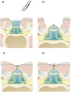
Open spina bifida repair technique. Step (a) shows untethering of the neural placode, followed by anatomical closure of the dura (b), myofascia (c) and skin (d). Reproduced with permission from Universitair Ziekenhuis Leuven, Belgium.
Figure 4
Figure 4
A case of myeloschisis. (a,b) Preoperative ultrasound findings of a relatively wide open lesion; (c) intraoperative images of the lesion prior to repair; (d) following closure of the dura and (e) application of a skin patch. Reproduced with permission from University College London Hospitals, London and Universitair Ziekenhuis Leuven, Belgium.
Figure 5
Figure 5
A case of myelomeningocele (same patient as Figure 2). (a) Preoperative findings of a covered lesion; (b) intraoperative images of the lesion prior to repair and (c) following skin closure. Reproduced with permission of University College London Hospitals and Universitair Ziekenhuis Leuven, Belgium.
Figure 6
Figure 6
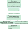
Outpatient postoperative pathway. FMU = fetal medicine unit; MRI = magnetic resonance imaging; SROM = spontaneous rupture of membranes; US = ultrasound scan
Figure 7
Figure 7
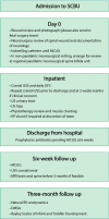
Neonatal protocol following open fetal surgery for spina bifida. DMSA = dimercaptosuccinic acid radionuclide scan; MCUG = micturating cystourethrogram; MRI = magnetic resonance imaging; OFC = occipito‐frontal circumference; SCBU = special care baby unit; USS = ultrasound scan; VP = ventriculoperitoneal
Figure 8
Figure 8
Examples of neurosurgical repair techniques reported in fetoscopic spina bifida repair. (a) Single layer repair (skin sutured); (b) double layer repair (subcutaneous patch and skin suture); (c) patch coverage; (d) double patch repair. Reproduced with permission of the Universitair Ziekenhuis Leuven, Belgium.

Similar articles

Cited by

References

    1. Khoshnood B, Loane M, de Walle H, Arriola L, Addor MC, Barisic I, et al. Long term trends in prevalence of neural tube defects in Europe: population based study. BMJ 2015;351:h5949. - PMC - PubMed
    1. Bowman RM, Mclone DG, Grant JA, Tomita T, Ito JA. Spina bifida outcome: a 25‐year prospective. Pediatr Neurosurg 2001;34:114–20. - PubMed
    1. de Jong TP, Chrzan R, Klijn AJ, Dik P. Treatment of the neurogenic bladder in spina bifida. Pediatr Nephrol 2008;23:889–96. - PMC - PubMed
    1. Velde Vande S, Van Biervliet S, Van Renterghem K, Van Laecke E, Hoebeke P, Van Winckel M. Achieving fecal continence in patients with spina bifida: a descriptive cohort study. J Urol 2007;178:2640–4. - PubMed
    1. Verhoef M, Barf HA, Vroege JA, Post MW, Van Asbeck FW, Gooskens RH, et al. Sex education, relationships, and sexuality in young adults with spina bifida. Arch Phys Med Rehabil 2005;86:979–87. - PubMed