. 2019 Nov 7:10:1316.
doi: 10.3389/fphar.2019.01316. eCollection 2019.

Involvement of Phosphatase and Tensin Homolog in Cyclin-Dependent Kinase 4/6 Inhibitor-Induced Blockade of Glioblastoma

Affiliations

Involvement of Phosphatase and Tensin Homolog in Cyclin-Dependent Kinase 4/6 Inhibitor-Induced Blockade of Glioblastoma

Songlin Liu et al. Front Pharmacol. .

Abstract

Dysregulation of retinoblastoma (Rb) signaling pathway have been established as a requirement for glioblastoma (GBM) initiation and progression, which suggests that blockade of CDK4/6-Rb signaling axis for GBM treatment. Palbociclib, a selective inhibitor of the cyclin-dependent kinases CDK4/6, has been applied for breast cancer treatment. However, its efficacy against glioblastoma has not been well clarified. Here, effects of CDK4/6 inhibitors on various kinds of GBM cell lines are investigated and the functional mechanisms are identified. Data showed that cells with diverse PTEN status respond to palbociclib differently. Gain-of-function and loss-of-function studies indicated that PTEN enhanced the sensitivity of GBM cells to palbociclib in vitro and in vivo, which was associated with suppressions of Akt and ERK signaling and independent of Rb signaling inhibition. Hence, our findings support that palbociclib selectively.

Keywords: CDK4/6 inhibitor; PTEN; glioblastoma; palbociclib; sensitivity.


Figures

Figure 1
Figure 1
Chemical structure of palbociclib (A) and ribociclib (B).
Figure 2
Figure 2
Effects of CDK4/6 inhibitor on various GBM cells with different PTEN status. (A, B). Cells were seeded in 96-well plate (3,000 cells/well) and treated with indicated concentrations of palbociclib (A) and ribociclib (B) for 48 h. Cell proliferation was assessed by MTT assay as described in Materials and Methods. Data are expressed as mean ± SD. (C) Palbociclib suppressed GBM cells in a time-dependent manner. Cells were prepared as described above and treated with palbociclib (0.5 μM) for indicated times. Cell proliferation was assessed by MTT assay as described in Materials and Methods. Data are expressed as mean ± SD. (D) Effects of palbociclib on colony formation in GBM cells with different PTEN status. Data are show as means ± SD (**P 0.01, t-test, n = 3). (E) PTEN expression profile at mRNA level in the tested GBM cell lines. (F) PTEN expression profile at protein level in the tested GBM cell lines.
Figure 3
Figure 3
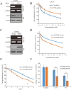
PTEN enhances the sensitivity of GBM cells to palbociclib. (A) Detection of PTEN expression at mRNA and protein levels in cells transfected with PTEN/control siRNA. Cell lysates/total RNA were collected and analyzed by western blot/RT-PCR assay as described in Materials and Methods. (B) PTEN knocking-down decreased the sensitivity of LN229 cells to palbociclib. Following PTEN/control siRNA transfection, cells were treated with palbociclib at indicated concentrations for 48 h. Cell proliferation was examined by MTT assay. Data are expressed as mean ± SD. (C) Detection of PTEN expression at mRNA and protein levels in isogenic U87MG cells. Cell lysates/total RNA were collected and examined by western blot/RT-PCR assay. (D) Expression of PTEN enhanced the efficacy of palbociclib against U87MG cells. Isogenic U87MG cells with or without PTEN were treated with palbociclib at indicated concentrations for 48 h, cell proliferation was assessed by MTT assay. Data are expressed as mean ± SD. (F) Time-course evaluation of palbociclib on isogenic U87MG cells. (E) Effects of palbociclib on colony formation of U87MG isogenic cells with different PTEN status. Data are shown as means ± SD (*P 0.05, **P 0.01, t-test, n = 3).
Figure 4
Figure 4
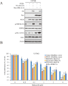
Palbociclib blocks ERK and Akt signaling in the presence of PTEN. (A) Effects of palbociclib on the levels of p-ERK, p-Akt, as well as p-Rb in LN229 cells pre-treated by PTEN/control siRNA. Following siRNA treatment, the cells were subjected to palbociclib treatment for 24 h. Cell lysates were collected and analyzed by western blot assay. (B) Quantification of the blots in (A) Data are shown as mean ± SD (*P 0.05, **P 0.01, one-way ANOVA, n = 3). (C) Effects of palbociclib on the levels of p-ERK, p-Akt, as well as p-Rb in U87MG isogenic cells. Cells were treated with palbociclib at indicated concentrations for 24 h. Cell lysates were collected and subjected to western blotting analysis with indicated antibodies. (D) Quantification of the blots in (C). Data are shown as mean ± SD (*P 0.05, **P 0.01, one-way ANOVA, n = 3).
Figure 5
Figure 5
ERK and Akt activation alleviates palbociclib-induced suppression of GBM cells. (A) Validation of the cell systems with active ERK or/and Akt activation. U87MG isogenic cells were transfected with ERK-CA or/and Akt-CA plasmids. Cell lysates were collected and examined by western blot assay with indicated antibodies. (B) Effects of palbociclib on cell proliferation in the cell systems constructed in (A) Following palbociclib treatment, cell proliferation was assessed by MTT assay as described in Materials and Methods. Data are expressed as mean ± SD (*P 0.05, **P 0.01, vs. control cells in each dose group, t-test, n = 3).
Figure 6
Figure 6
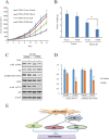
Palbociclib selectively blocks PTEN-expressed U87MG cells in vivo. (A, B) U87MG isogenic cells were inoculated subcutaneously; mice were treated with palbociclib as described in Materials and Methods. Tumor volumes (A) and tumor weight (B) were examined throughout the experiment. Data are shown as mean ± SD (*P 0.05, **P 0.01, t-test, eight mice/group). (C) Effects of palbociclib on the levels of p-ERK, p-Akt, as well as p-Rb in U87MG-Vector/PTEN tumors. Following treatment, tumor tissues were collected. The lysates were analyzed by western blot assay with indicated antibodies. (D) Quantification of the blots in (C). Data are shown as mean ± SD (**P 0.01, vs. vehicle treatment, t-test, n = 3). (E) A schematic diagram

Similar articles

Cited by

References

    1. Bollard J., Miguela V., Ruiz de Galarreta M., Venkatesh A., Bian C. B., Roberto M. P., et al. (2016). Palbociclib (PD-0332991), a selective CDK4/6 inhibitor, restricts tumour growth in preclinical models of hepatocellular carcinoma. Gut. 66, 1286–1296. 10.1136/gutjnl-2016-312268 - DOI - PMC - PubMed
    1. Brennan C. W., Verhaak R. G., McKenna A., Campos B., Noushmehr H., Salama S. R., et al. (2013). The somatic genomic landscape of glioblastoma. Cell 155, 462–477. 10.1016/j.cell.2013.09.034 - DOI - PMC - PubMed
    1. Cen L., Carlson B. L., Schroeder M. A., Ostrem J. L., Kitange G. J., Mladek A. C., et al. (2012). p16-Cdk4-Rb axis controls sensitivity to a cyclin-dependent kinase inhibitor PD0332991 in glioblastoma xenograft cells. Neuro-oncology 14, 870–881. 10.1093/neuonc/nos114 - DOI - PMC - PubMed
    1. Chen L., Pan J. (2017). Dual cyclin-dependent kinase 4/6 inhibition by PD-0332991 induces apoptosis and senescence in oesophageal squamous cell carcinoma cells. Br. J. Pharmacol. 174, 2427–2443. 10.1111/bph.13836 - DOI - PMC - PubMed
    1. Chenn A., Walsh C. A. (2002). Regulation of cerebral cortical size by control of cell cycle exit in neural precursors. Science 297, 365–369. 10.1126/science.1074192 - DOI - PubMed