. 2019 Nov 15;4(22):19913-19924.
doi: 10.1021/acsomega.9b02824. eCollection 2019 Nov 26.

Unique Structural Features of Mule Deer Prion Protein Provide Insights into Chronic Wasting Disease

Affiliations

Unique Structural Features of Mule Deer Prion Protein Provide Insights into Chronic Wasting Disease

Urška Slapšak et al. ACS Omega. .

Abstract

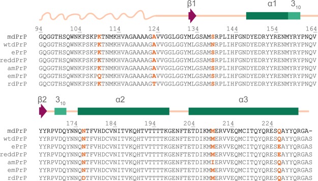
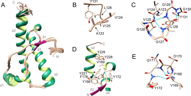
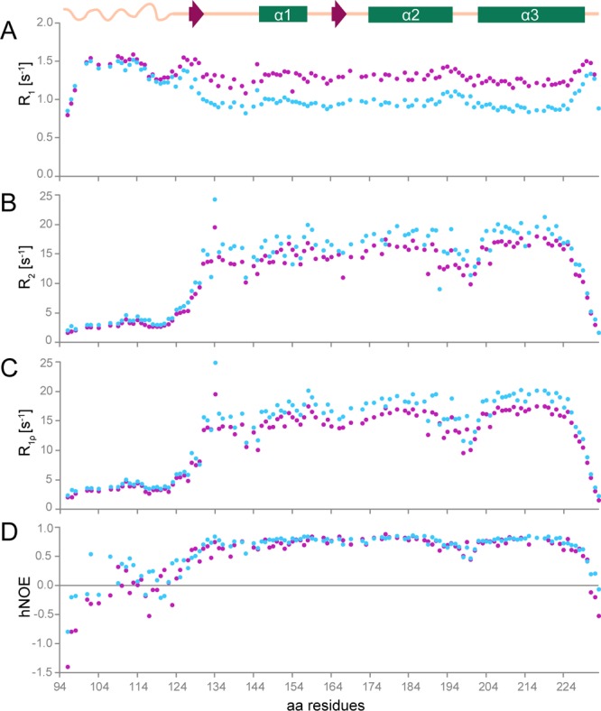
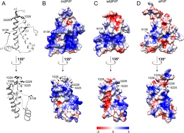
Chronic wasting disease (CWD) is a highly infectious prion disease of cervids. Accumulation of prions, the disease-specific structural conformers of the cellular prion protein (PrPC), in the central nervous system, is the key pathological event of the disorder. The analysis of cervid PrPC sequences revealed the existence of polymorphism at position 226, in which deer PrP contains glutamine (Q), whereas elk PrP contains glutamate (E). The effects of this polymorphism on CWD are still unknown. We determined the high-resolution nuclear magnetic resonance structure of the mule deer prion protein that was compared to previously published PrP structures of elk and white-tailed deer. We found that the polymorphism Q226E could influence the long-range intramolecular interactions and packing of the β2-α2 loop and the C-terminus of the α3 helix of cervid PrP structures. This solvent-accessible epitope is believed to be involved in prion conversion. Additional differences were observed at the beginning of the well-defined C-terminus domain, in the α2-α3 region, and in its interactions with the α1 helix. Here, we highlight the importance of the PrP structure in prion susceptibility and how single amino acid differences might influence the overall protein folding.


Conflict of interest statement

The authors declare no competing financial interest.

Figures

Figure 1
Figure 1
Sequence alignment of PrPs of cervid subspecies with confirmed CWD. Amino acid variants are marked with orange color. Residue numbering is based on the mdPrP amino acid sequence. Secondary structural elements are summarized based on the mdPrP structural model presented in this article, with the α-helices of mdPrP denoted by green rectangles, 310-helices by light green rectangles, β-strands by magenta arrows, flexible N-terminal tail by a curved line, and linkers between the secondary structure elements by straight lines, both lines colored champagne pink.
Figure 2
Figure 2
15N-HSQC spectrum of mdPrP with the amino acid assignment. Cross-peaks of the side chains of Asn, Gln, and Trp are not marked.
Figure 3
Figure 3
Structure of mdPrP. (A) Ensemble of 20 lowest energy structures of mdPrP (residues form Ala123 to Ala233). α-Helices and 310-helix are colored green, β-sheets are colored magenta, and loops are colored champagne pink. (B) Well-defined region between residues Ala123 and Tyr131. (C) Residues from Ala123 to Tyr131 involved in the formation of α-helical turn (Val125–Leu128) and γ-turn (Leu128–Gly130). (D) Hydrophobic pocket in the proximity of the β2−α2 loop and the C-terminus of the α3 helix. (E) 310-Helix from residues Pro168 to Tyr172 inside the β2−α2 loop. Residues are presented as sticks in champagne pink and the hydrogen bonds in panels (C,E) are shown as dashed lines in cyan.
Figure 4
Figure 4
15N amide backbone relaxation rates and hNOE of mdPrP. (A) 15N longitudinal (R1 = 1/T1), (B) transverse (R2 = 1/T2), (C) spin–lattice relaxation rates in the rotation frame (R = 1/T), and (D) hNOE at 298 K at a magnetic field of 14.1 (magenta) and 18.8 T (blue). A schematic presentation of the secondary structure elements of mdPrP is at the top of the figure. For clarity, error bars are not shown here as they are within the size of the data points in the above graphics but are presented in the Supporting Information in Figure S1.
Figure 5
Figure 5
Comparison of mdPrP, wtdPrP, and ePrP structures. (A) Superposition of well-defined C-terminus domains from amino acids Ala123–Ala233 of mdPrP (green), wtdPrP (orange), and ePrP (magenta). The selected residues are presented as ball-and-stick and colored in champagne pink with marked heteroatoms. (B) Structural diversity at the end of the α3 helix and the β2−α2 loop. (C) Spatial orientation of residues in the proximity of the α2−α3 loop with marked distances. Selected distances among residues are indicated with dashed lines and small letters (see Table 2 for distance information). (D) Structural differences in orientations at the α1 helix with respect to the α2−α3 V-shaped skeleton.
Figure 6
Figure 6
Local rmsd values for backbone atoms per residue (from 128 to 228) of mdPrP (green) and ePrP (magenta) with respect to the wtdPrP structure that was determined by X-ray. Standard deviations are reported for the ensemble of 20 lowest energy structures of mdPrP and ePrP.
Figure 7
Figure 7
Solvent accessibility of selected residues that belong to the α2 and α3 helices. Hatched and dotted lines at 20 and 50% indicate the limits of amino acid residue accessibility to solvents (>50%) or burial in solvent-inaccessible regions (<20%). Standard deviations are reported for the ensemble of 20 lowest energy structures of mdPrP and ePrP that have been determined by NMR.
Figure 8
Figure 8
Electrostatic surface potential of the three cervid PrPs. (A) Ribbon presentation of the mdPrP backbone orientation used in panels (B–D). Residues Ser138, Ser225, Gln226, Tyr228, and Tyr229 are presented as ball-and-stick and colored black. Electrostatic surface potentials of (B) mdPrP, (C) wtdPrP, and (D) ePrP. Regions of positive and negative charges are depicted from blue to red according to the presented charge legend. Orientation of structures is preserved in all panels. The lower set of structures is rotated by 135°.

References

    1. Belay E. D.; Maddox R. A.; Williams E. S.; Miller M. W.; Gambetti P.; Schonberger L. B. Chronic wasting disease and potential transmission to humans. Emerging Infect. Dis. 2004, 10, 977.10.3201/eid1006.031082. - DOI - PMC - PubMed
    1. Haley N. J.; Hoover E. A. Chronic wasting disease of cervids: current knowledge and future perspectives. Annu. Rev. Anim. Biosci. 2015, 3, 305–325. 10.1146/annurev-animal-022114-111001. - DOI - PubMed
    1. Williams E. S.; Young S. Chronic wasting disease of captive mule deer: A spongiform encephalopathy 1. J. Wildl. Dis. 1980, 16, 89–98. 10.7589/0090-3558-16.1.89. - DOI - PubMed
    1. Williams E. S.; Young S. Spongiform encephalopathy of rocky mountain elk 1. J. Wildl. Dis. 1982, 18, 465–471. 10.7589/0090-3558-18.4.465. - DOI - PubMed
    1. Spraker T. R.; Miller M. W.; Williams E. S.; Getzy D. M.; Adrian W. J.; Schoonveld G. G.; Spowart R. A.; O’Rourke K. I.; Miller J. M.; Merz P. A. Spongiform encephalopathy in free-ranging mule deer (Odocoileus hemionus), white-tailed deer (Odocoileus virginianus) and Rocky Mountain elk (Cervus elaphus nelsoni) in northcentral Colorado. J. Wildl. Dis. 1997, 33, 1–6. 10.7589/0090-3558-33.1.1. - DOI - PubMed