

Hemoglobin Video Imaging Provides Novel In Vivo High-Resolution Imaging and Quantification of Human Aqueous Outflow in Patients with Glaucoma
- PMID: 31788668
- PMCID: PMC6876656
- DOI: 10.1016/j.ogla.2019.04.001
Hemoglobin Video Imaging Provides Novel In Vivo High-Resolution Imaging and Quantification of Human Aqueous Outflow in Patients with Glaucoma
Abstract
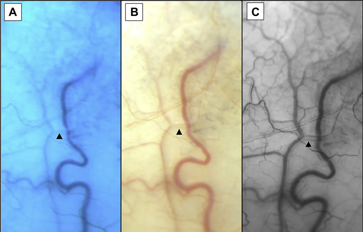
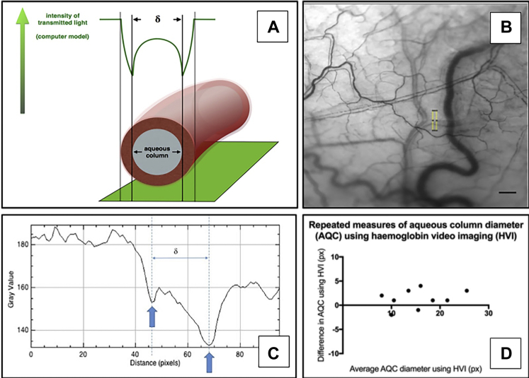
Purpose: Noninvasive, detailed measurement of the dynamics of human aqueous outflow is difficult to achieve with currently available clinical tools. We used hemoglobin video imaging (HVI) to develop a technique to image and quantify human aqueous outflow noninvasively and in real time.
Design: A prospective observational study to describe characteristics of aqueous veins and a pilot prospective interventional feasibility study to develop quantification parameters.
Participants: Patients were recruited from the Cambridge University Hospitals NHS Foundation Trust Glaucoma clinic. The observational study included 30 eyes, and the pilot interventional feasibility study was performed on 8 eyes undergoing selective laser trabeculoplasty (SLT). Our SLT protocol also included the installation of pilocarpine and apraclonidine eye drops.
Methods: Participants underwent HVI alongside their usual clinic visit.
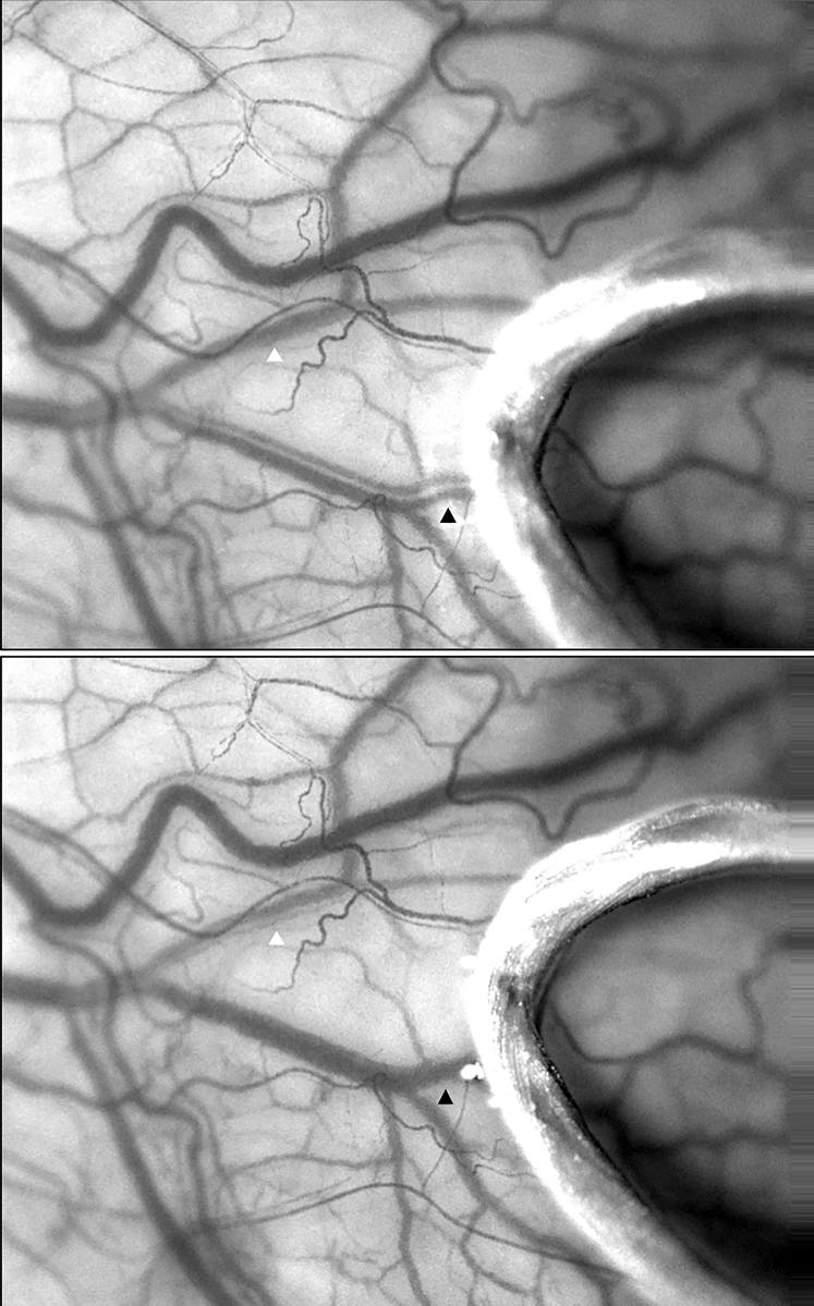
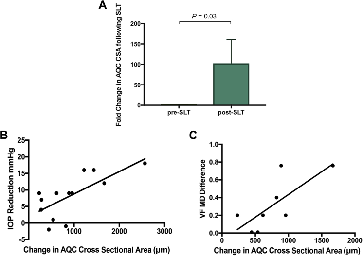
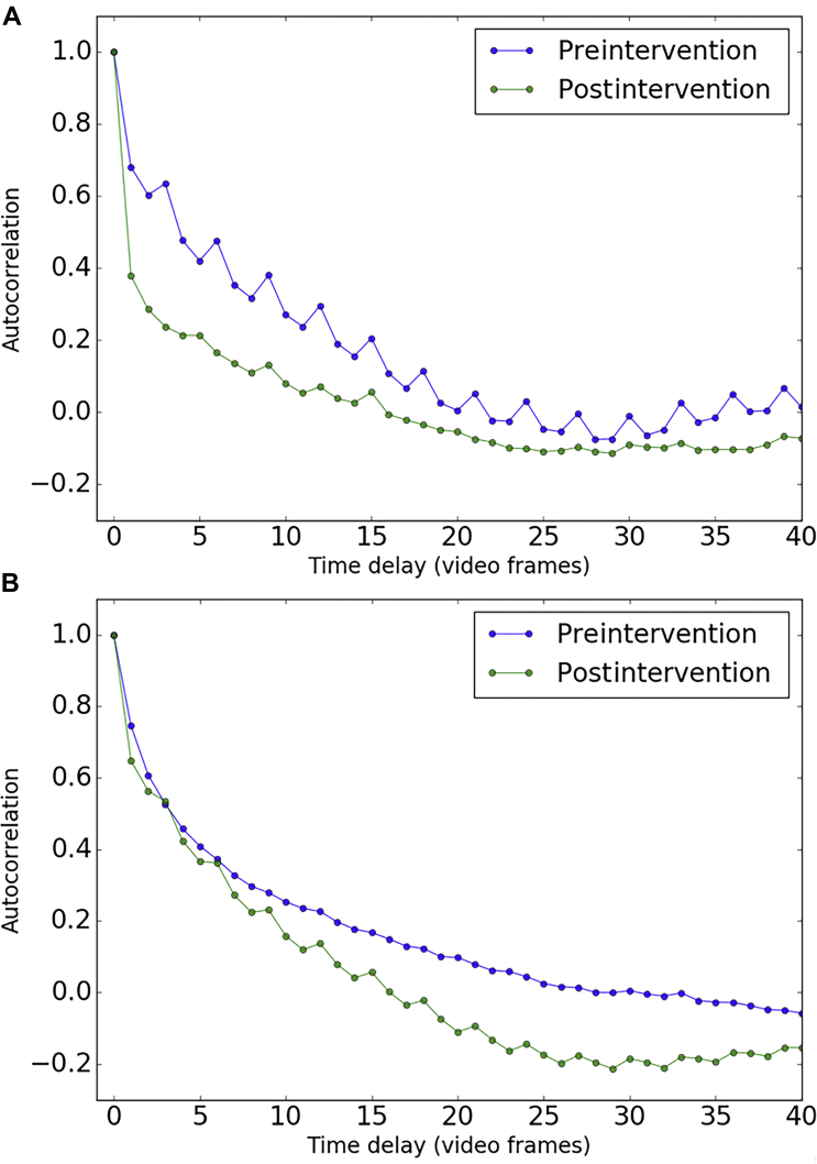
Main outcome measures: The change in cross-sectional area (CSA) of the aqueous column within episcleral veins was correlated with intraocular pressure (IOP) reduction and change in visual field mean deviation (MD) before and after intervention. Fluctuations in contrast and pixel intensity of red blood cells in an aqueous vein were calculated to compare the flow rate before and after intervention using autocorrelation analysis.
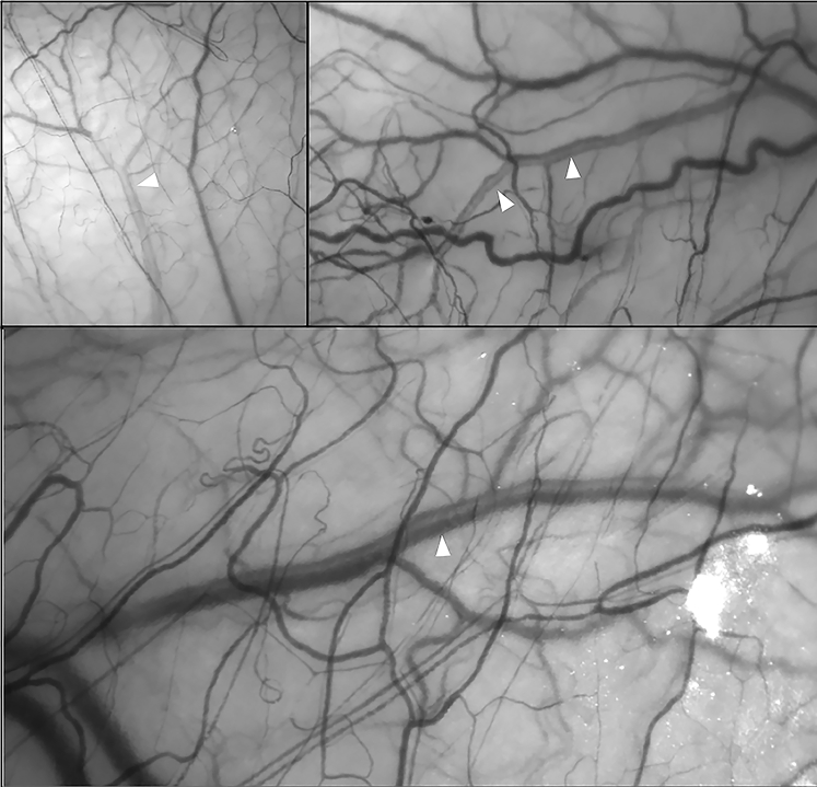
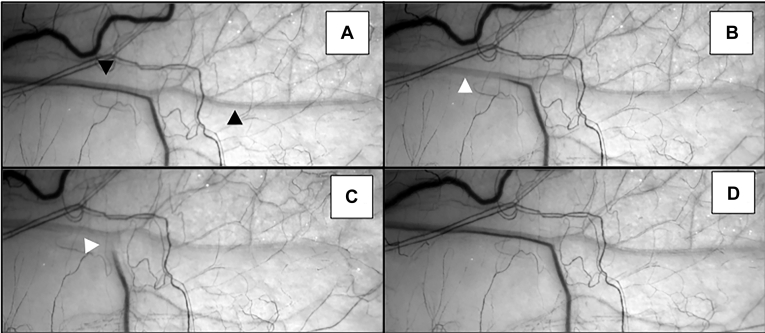
Results: Hemoglobin video imaging enables the direct observation of aqueous flow into the vascular system. Aqueous is seen to centralize within a laminar venous column. Flow is pulsatile, and fluctuations of flow through globe pressure or compression of the aqueous vein are observed. There was a significant increase in the aqueous column after the administration of our SLT protocol (n = 13; P < 0.05). This correlated with the degree of IOP reduction (n = 13; Pearson's correlation coefficient 0.7; P = 0.007) and the improvement in MD observed postintervention (n = 8; Pearson's correlation coefficient 0.75; P = 0.03). Autocorrelation analysis demonstrated a faster rate of decay in an aqueous vein after intervention, indicating an increase in flow rate.
Conclusions: Hemoglobin video imaging can be incorporated into a routine clinic slit-lamp examination to allow a detailed assessment and quantification of aqueous outflow in real time. It has the potential to be used to help target therapeutic interventions to improve aqueous outflow and further advance our understanding of aqueous outflow dysregulation in the pathogenesis of glaucoma.
Keywords: CSA, cross-sectional area; HVI, hemoglobin video imaging; IOP, intraocular pressure; MD, mean deviation; MIGS, minimally invasive glaucoma surgery; SLT, selective laser trabeculoplasty.
© 2019 by the American Academy of OphthalmologyThis is an open access article under the CC BY license (
Figures
References
-
- Johnstone M.A. The aqueous outflow system as a mechanical pump: evidence from examination of tissue and aqueous movement in human and non-human primates. J Glaucoma. 2004;13:421–438. - PubMed
Publication types
MeSH terms
Substances
Grants and funding
LinkOut - more resources
Full Text Sources
