

Andrographolide sodium bisulfate attenuates UV‑induced photo‑damage by activating the keap1/Nrf2 pathway and downregulating the NF‑κB pathway in HaCaT keratinocytes
- PMID: 31789424
- PMCID: PMC6984792
- DOI: 10.3892/ijmm.2019.4415
Andrographolide sodium bisulfate attenuates UV‑induced photo‑damage by activating the keap1/Nrf2 pathway and downregulating the NF‑κB pathway in HaCaT keratinocytes
Abstract
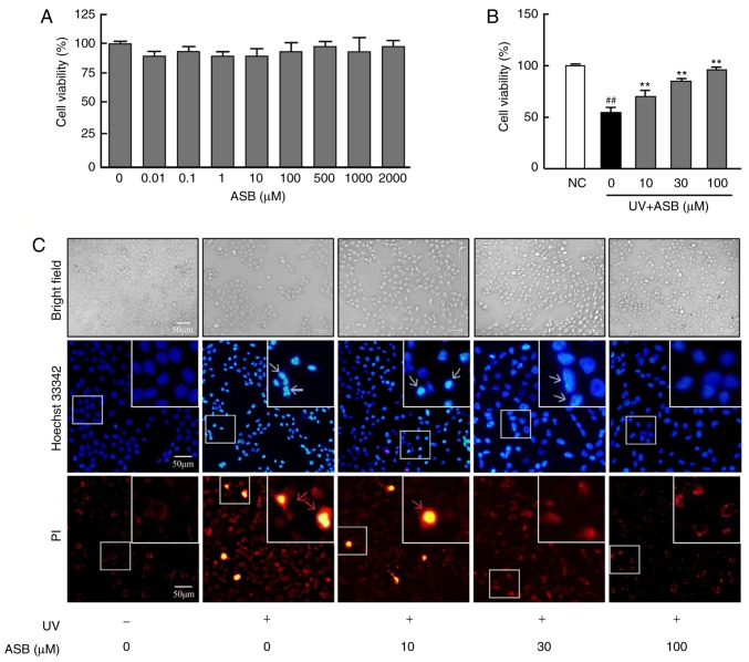
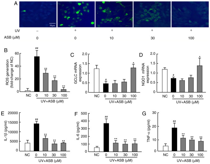
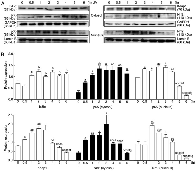
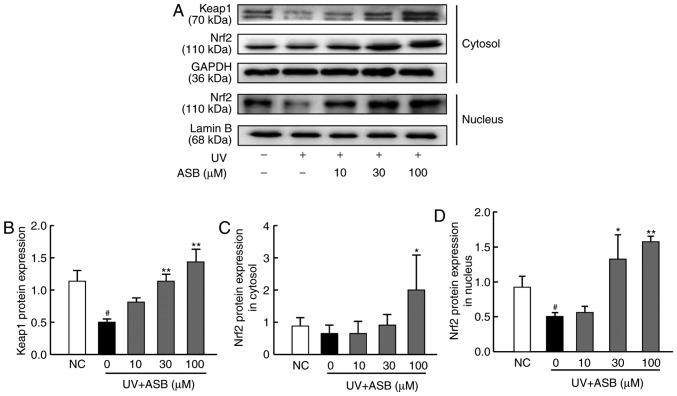
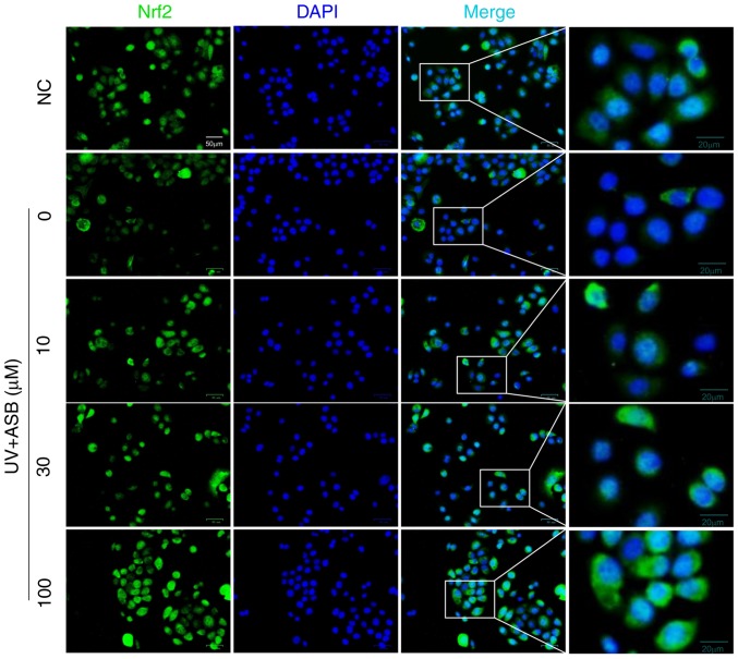
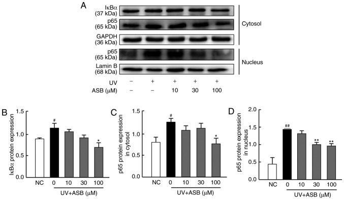
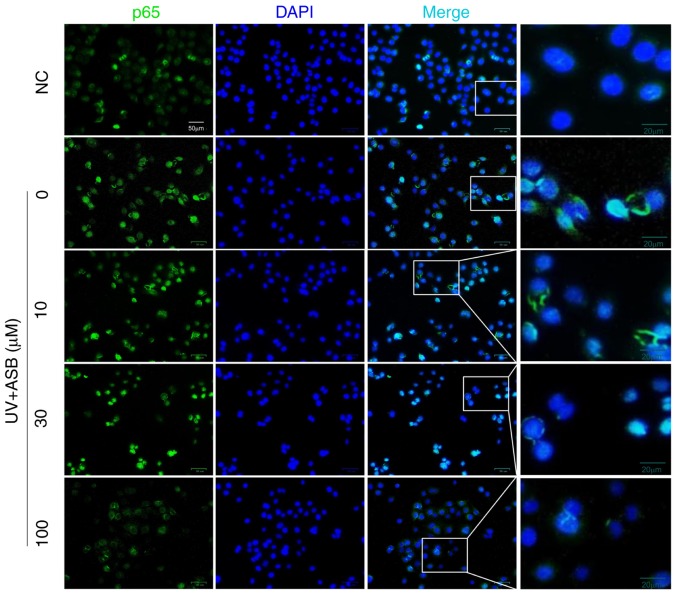
Oxidative and inflammatory damage has been suggested to play important roles in the pathogenesis of skin photoaging. Andrographolide sodium bisulfate (ASB) is a soluble derivative of andrographolide and has known antioxidant and anti‑inflammatory properties. In the present study, cellular experiments were designed to investigate the molecular mechanisms underlying the effect of ASB in relieving ultraviolet (UV)‑induced photo‑damage. Following ASB pretreatment and UV irradiation, the apoptosis and necrosis of HaCaT cells were investigated by Hoechst 33342/propidium iodide staining. Reactive oxygen species (ROS) production was investigated using a DCFH‑DA fluorescence probe. Furthermore, the protein expression levels of p65, NF‑κB inhibitor‑α, nuclear factor E2‑related factor 2 (Nrf2) and kelch‑like ECH‑associated protein 1 (keap1) were measured via western blotting and immunofluorescence analyses. Furthermore, NF‑κB‑mediated cytokines were assessed by ELISA, and Nrf2‑mediated genes were detected by reverse transcription‑quantitative PCR. Pretreatment with ASB markedly increased cell viability, decreased cell apoptosis and decreased UV‑induced excess ROS levels. In addition, ASB activated the production of Nrf2 and increased the mRNA expression levels of glutamate‑cysteine ligase catalytic subunit and NAD(P)H quinone oxidoreductase 1, while ASB downregulated the protein expression of p65 and decreased the production of interleukin (IL)‑1β, IL‑6 and tumor necrosis factor‑α. These results suggested that ASB attenuates UV‑induced photo‑damage by activating the keap1/Nrf2 pathway and downregulating the NF‑κB pathway in HaCaT keratinocytes.
Figures

Similar articles
-
Resveratrol protects human keratinocytes HaCaT cells from UVA-induced oxidative stress damage by downregulating Keap1 expression.Eur J Pharmacol. 2011 Jan 10;650(1):130-7. doi: 10.1016/j.ejphar.2010.10.009. Epub 2010 Oct 14. Eur J Pharmacol. 2011. PMID: 20951123
-
Lycium barbarum polysaccharides attenuate cardiovascular oxidative stress injury by enhancing the Keap1/Nrf2 signaling pathway in exhaustive exercise rats.Mol Med Rep. 2021 Sep;24(3):643. doi: 10.3892/mmr.2021.12282. Epub 2021 Jul 19. Mol Med Rep. 2021. PMID: 34278476
-
Protective roles of NRF2 signaling pathway in cobalt chloride-induced hypoxic cytotoxicity in human HaCaT keratinocytes.Toxicol Appl Pharmacol. 2018 Sep 15;355:189-197. doi: 10.1016/j.taap.2018.06.030. Epub 2018 Jun 30. Toxicol Appl Pharmacol. 2018. PMID: 29966676
-
Targeting NRF2 for Improved Skin Barrier Function and Photoprotection: Focus on the Achiote-Derived Apocarotenoid Bixin.Nutrients. 2017 Dec 18;9(12):1371. doi: 10.3390/nu9121371. Nutrients. 2017. PMID: 29258247 Free PMC article. Review.
-
Roles of the KEAP1-NRF2 system in mammalian skin exposed to UV radiation.Toxicol Appl Pharmacol. 2018 Dec 1;360:69-77. doi: 10.1016/j.taap.2018.09.038. Epub 2018 Sep 28. Toxicol Appl Pharmacol. 2018. PMID: 30268578 Review.
Cited by
-
Skincare Benefits of a Postbiotic Ferment Produced Through Djon Djon Mushroom Fermentation by Saccharomyces.J Cosmet Dermatol. 2025 Feb;24(2):e70067. doi: 10.1111/jocd.70067. J Cosmet Dermatol. 2025. PMID: 39968713 Free PMC article.
-
Andrographis paniculata and Its Bioactive Diterpenoids Against Inflammation and Oxidative Stress in Keratinocytes.Antioxidants (Basel). 2020 Jun 17;9(6):530. doi: 10.3390/antiox9060530. Antioxidants (Basel). 2020. PMID: 32560449 Free PMC article.
-
Bromophenol Bis (2,3,6-Tribromo-4,5-dihydroxybenzyl) Ether Protects HaCaT Skin Cells from Oxidative Damage via Nrf2-Mediated Pathways.Antioxidants (Basel). 2021 Sep 9;10(9):1436. doi: 10.3390/antiox10091436. Antioxidants (Basel). 2021. PMID: 34573068 Free PMC article.
-
Dysregulated CREB3 cleavage at the nuclear membrane induces karyoptosis-mediated cell death.Exp Mol Med. 2024 Mar;56(3):686-699. doi: 10.1038/s12276-024-01195-1. Epub 2024 Mar 13. Exp Mol Med. 2024. PMID: 38480902 Free PMC article.
-
Andrographolide Mitigates Inflammation and Reverses UVB-Induced Metabolic Reprogramming in HaCaT Cells.Int J Mol Sci. 2025 Jul 6;26(13):6508. doi: 10.3390/ijms26136508. Int J Mol Sci. 2025. PMID: 40650284 Free PMC article.
References
-
- Puizina-Ivić N. Skin aging. Acta Dermatovenerol Alp Pannonica Adriat. 2008;17:47–54. - PubMed
MeSH terms
Substances
LinkOut - more resources
Full Text Sources
Research Materials
