

Multimodal, in Situ Imaging of Ex Vivo Human Skin Reveals Decrease of Cholesterol Sulfate in the Neoepithelium during Acute Wound Healing
- PMID: 31789498
- PMCID: PMC8341291
- DOI: 10.1021/acs.analchem.9b04542
Multimodal, in Situ Imaging of Ex Vivo Human Skin Reveals Decrease of Cholesterol Sulfate in the Neoepithelium during Acute Wound Healing
Abstract
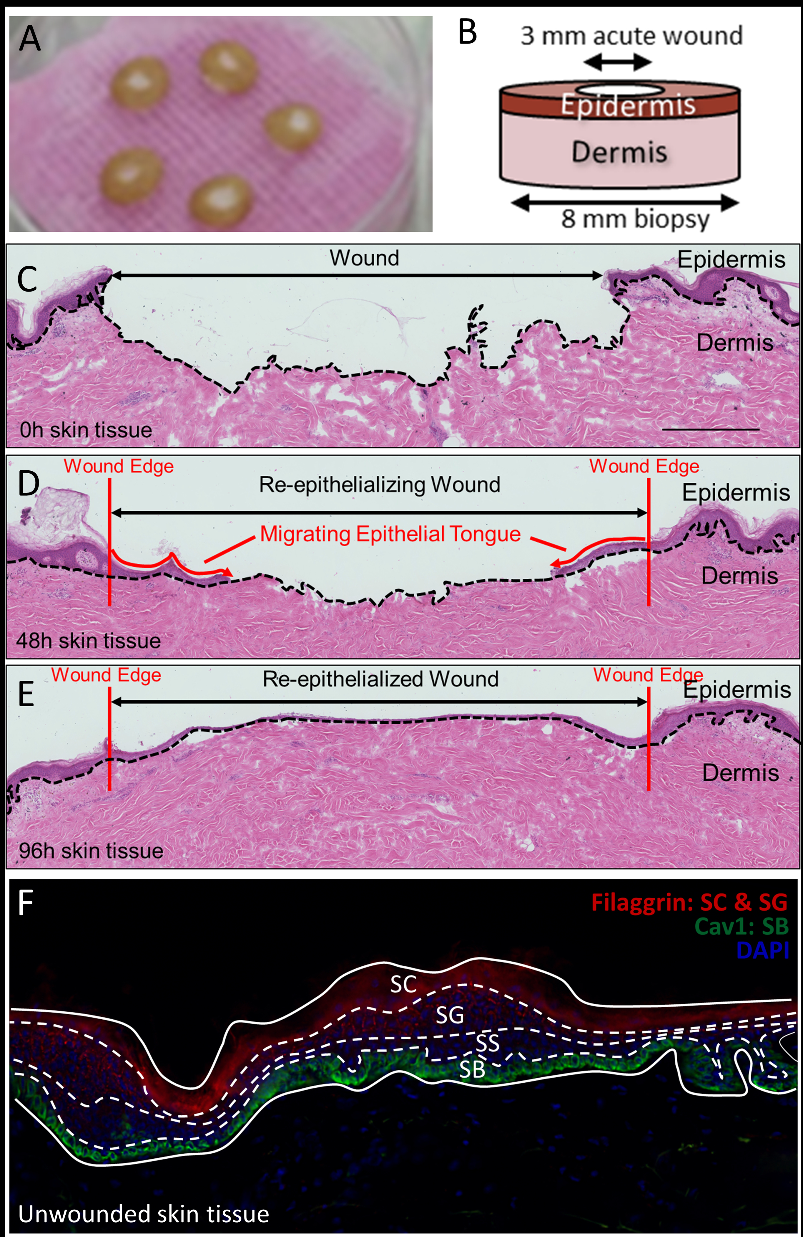
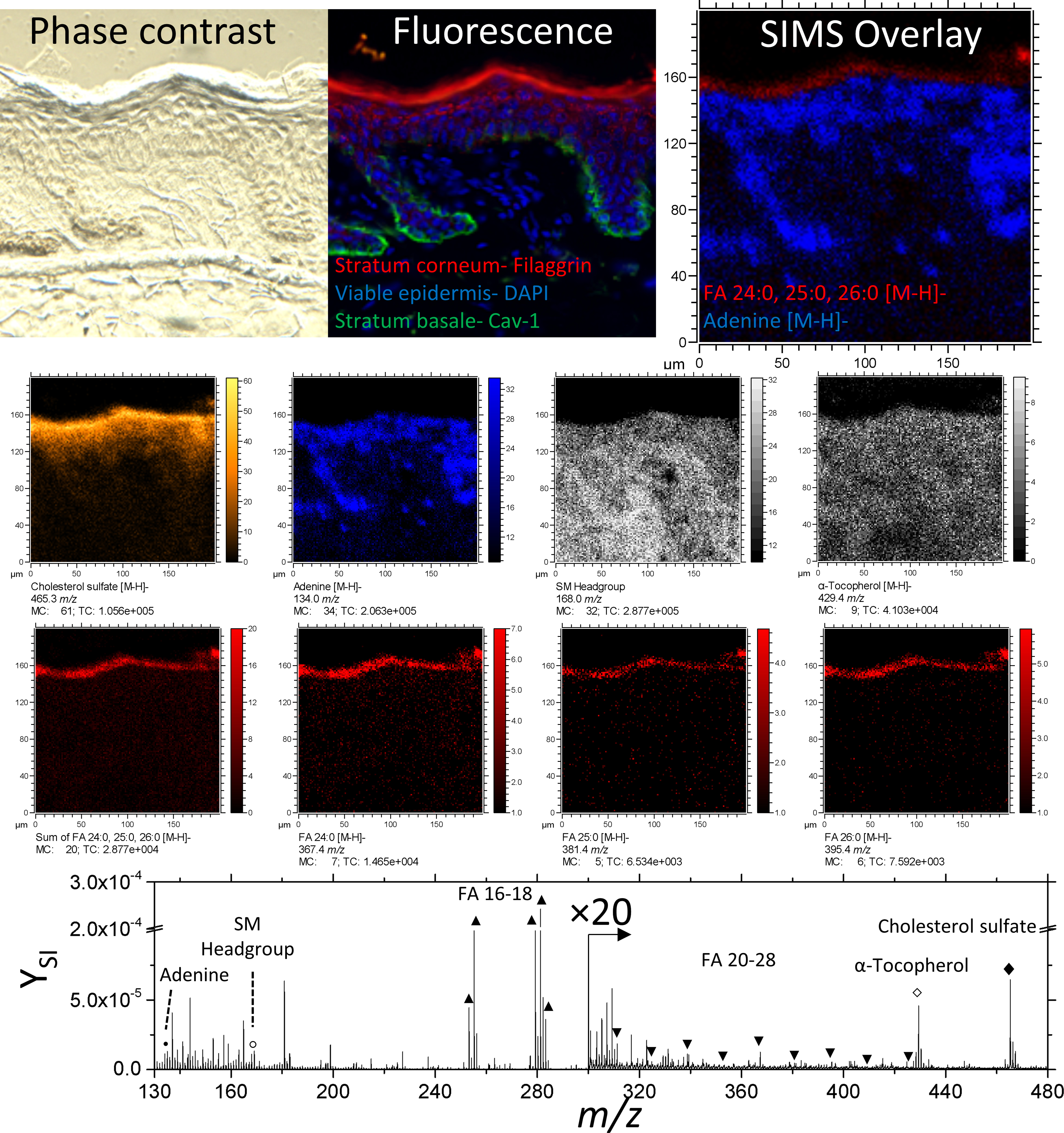
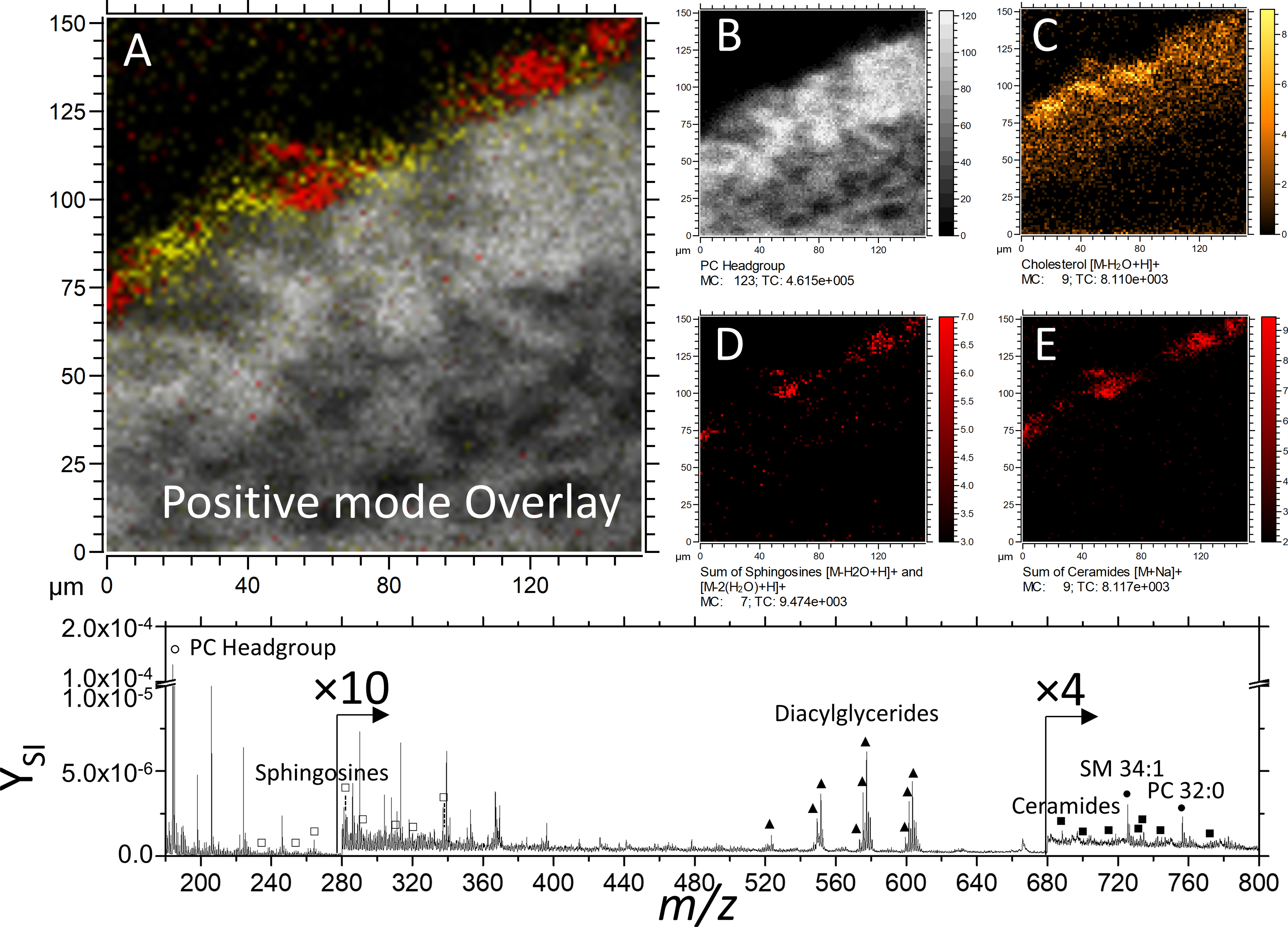
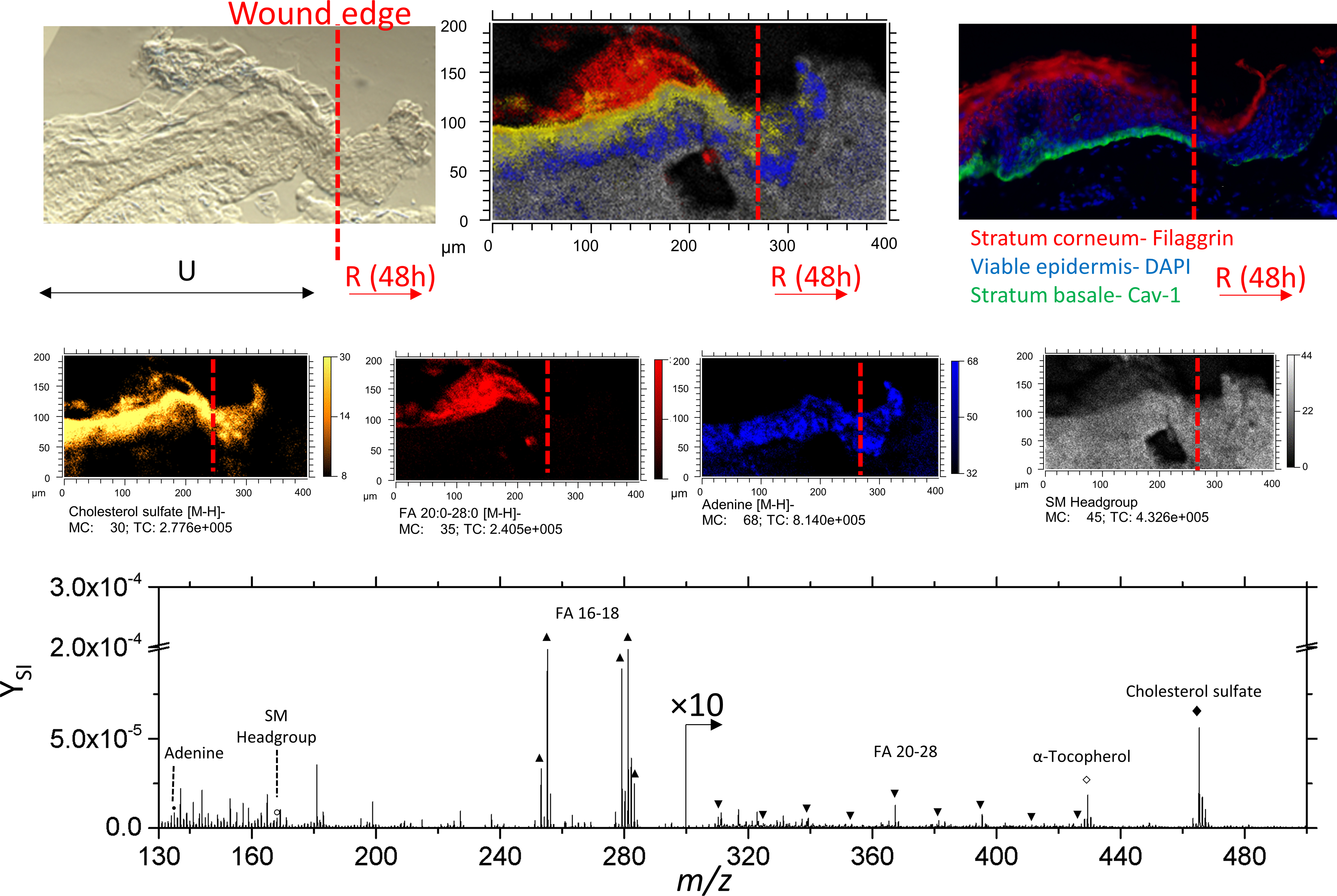
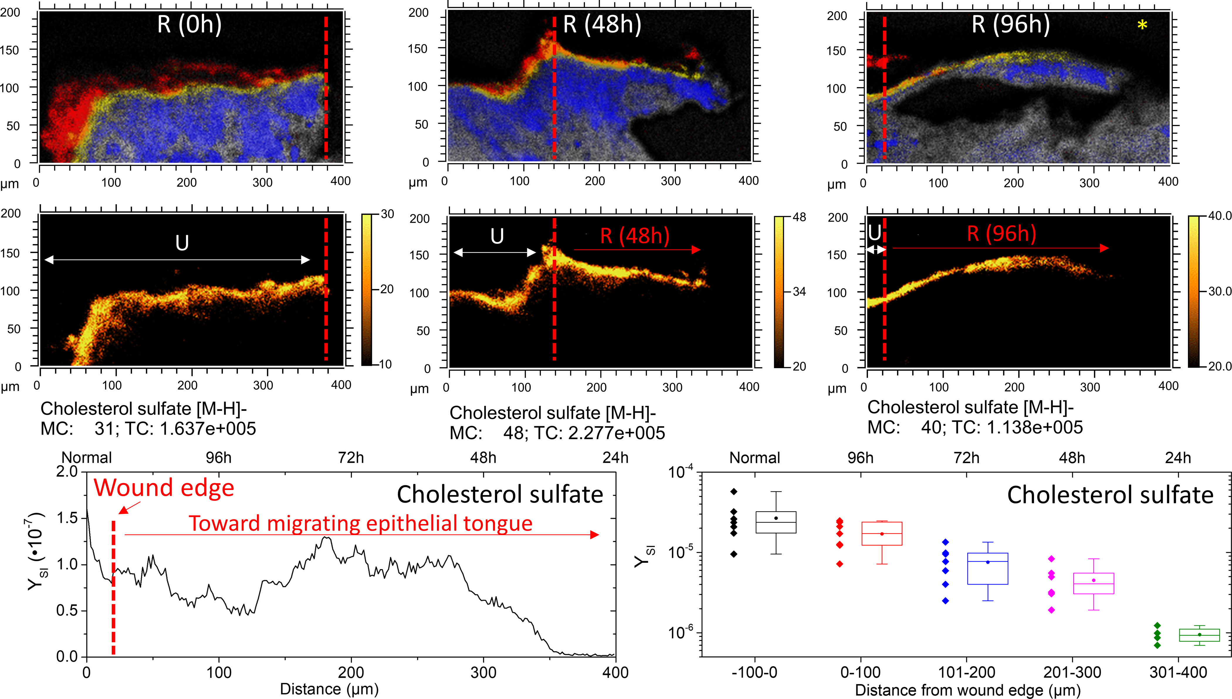
Skin repair is a significant aspect of human health. While the makeup of healthy stratum corneum and epidermis is generally understood, the mobilization of molecular components during skin repair remains largely unknown. In the present work, we utilize multimodal, in situ, mass spectrometry, and immunofluorescence imaging for the characterization of newly formed epidermis, following an initial acute wound for the first 96 h of epithelization. In particular, TOF-SIMS and confirmatory MALDI FT-ICR MS (/MS) analysis permitted the mapping of several lipid classes, including phospholipids, neutral lipids, cholesterol, ceramides, and free fatty acids. Endogenous lipid species were localized in discrete epidermal skin layers, including the stratum corneum (SC), stratum granulosum (SG), stratum basale (SB), and dermis. Experiments revealed that healthy re-epithelializing skin is characterized by diminished cholesterol sulfate signal along the stratum corneum toward the migrating epithelial tongue. The spatial distribution and relative abundances of cholesterol sulfate are reported and correlated with the healing time. The multimodal imaging approach enabled in situ high-confidence chemical mapping based on accurate mass and fragmentation pattern of molecular components. The use of postanalysis immunofluorescence imaging from the same tissue confirmed the localization of endogenous lipid species and Filaggrin and Cav-1 proteins at high spatial resolution (approximately a few microns).
Conflict of interest statement
The authors declare no competing financial interest.
Figures
Similar articles
-
Stratum corneum lipids in disorders of cornification. Steroid sulfatase and cholesterol sulfate in normal desquamation and the pathogenesis of recessive X-linked ichthyosis.J Clin Invest. 1984 Oct;74(4):1414-21. doi: 10.1172/JCI111552. J Clin Invest. 1984. PMID: 6592175 Free PMC article.
-
3D Molecular Imaging of Stratum Corneum by Mass Spectrometry Suggests Distinct Distribution of Cholesteryl Esters Compared to Other Skin Lipids.Int J Mol Sci. 2022 Nov 9;23(22):13799. doi: 10.3390/ijms232213799. Int J Mol Sci. 2022. PMID: 36430276 Free PMC article.
-
Skin diseases associated with the depletion of stratum corneum lipids and stratum corneum lipid substitution therapy.Skin Pharmacol Physiol. 2015;28(1):42-55. doi: 10.1159/000360009. Epub 2014 Aug 29. Skin Pharmacol Physiol. 2015. PMID: 25196193 Review.
-
Cholesterol sulfate and calcium affect stratum corneum lipid organization over a wide temperature range.J Lipid Res. 1999 Dec;40(12):2303-12. J Lipid Res. 1999. PMID: 10588956
-
Stratum Corneum Lipids: Their Role for the Skin Barrier Function in Healthy Subjects and Atopic Dermatitis Patients.Curr Probl Dermatol. 2016;49:8-26. doi: 10.1159/000441540. Epub 2016 Feb 4. Curr Probl Dermatol. 2016. PMID: 26844894 Review.
Cited by
-
Upregulation of Caveolae-Associated Proteins in Lesional Samples of Hidradenitis Suppurativa: A Case Series Study.JID Innov. 2023 Aug 14;3(6):100223. doi: 10.1016/j.xjidi.2023.100223. eCollection 2023 Nov. JID Innov. 2023. PMID: 37731470 Free PMC article.
-
Downregulation of Caveolae-Associated Proteins in Psoriasis: A Case Series Study.JID Innov. 2024 Feb 1;4(2):100265. doi: 10.1016/j.xjidi.2024.100265. eCollection 2024 Mar. JID Innov. 2024. PMID: 38445230 Free PMC article.
-
Glucocorticoid-mediated induction of caveolin-1 disrupts cytoskeletal organization, inhibits cell migration and re-epithelialization of non-healing wounds.Commun Biol. 2021 Jun 18;4(1):757. doi: 10.1038/s42003-021-02298-5. Commun Biol. 2021. PMID: 34145387 Free PMC article.
-
Direct Sampling Mass Spectrometry Analysis for the Assessment of Wounds: A Systematic Review.Int Wound J. 2025 Apr;22(4):e70158. doi: 10.1111/iwj.70158. Int Wound J. 2025. PMID: 40129114 Free PMC article.
-
Three-dimensional (3D) imaging of lipids in skin tissues with infrared matrix-assisted laser desorption electrospray ionization (MALDESI) mass spectrometry.Anal Bioanal Chem. 2021 Apr;413(10):2793-2801. doi: 10.1007/s00216-020-03105-6. Epub 2021 Jan 2. Anal Bioanal Chem. 2021. PMID: 33388847 Free PMC article.
References
-
- Feingold KR, Thematic review series: Skin Lipids. The role of epidermal lipids in cutaneous permeability barrier homeostasis. Journal of Lipid Research 2007, 48 (12), 2531–2546. - PubMed
-
- Armstrong DG; Boulton AJM; Bus SA, Diabetic Foot Ulcers and Their Recurrence. New England Journal of Medicine 2017, 376 (24), 2367–2375. - PubMed
-
- Nussbaum SR; Carter MJ; Fife CE; DaVanzo J; Haught R; Nusgart M; Cartwright D, An Economic Evaluation of the Impact, Cost, and Medicare Policy Implications of Chronic Nonhealing Wounds. Value Health 2018, 21 (1), 27–32. - PubMed
Publication types
MeSH terms
Substances
Grants and funding
LinkOut - more resources
Full Text Sources
