. 2020 Jan/Feb;44(1):43-46.
doi: 10.1097/RCT.0000000000000944.

Hippocampal Sulcus Remnant: Common Finding in Nonelderly Adults on Ultra-High-Resolution 7T Magnetic Resonance Imaging

Affiliations

Hippocampal Sulcus Remnant: Common Finding in Nonelderly Adults on Ultra-High-Resolution 7T Magnetic Resonance Imaging

Koji Yamashita et al. J Comput Assist Tomogr. 2020 Jan/Feb.

Abstract

Objective: The objective of this study was to investigate the frequency of hippocampal sulcus remnants (HSRs) in nonelderly adults using ultra-high-resolution 7T magnetic resonance (MR) images and their imaging features.

Methods: A total of 33 healthy adults underwent 7T MR, and multiplanar images of 66 temporal lobes were reviewed independently by 2 neuroradiologists. The detectability of the HSR was calculated. In addition, the interobserver agreement on the rating scale was evaluated using the κ statistic.

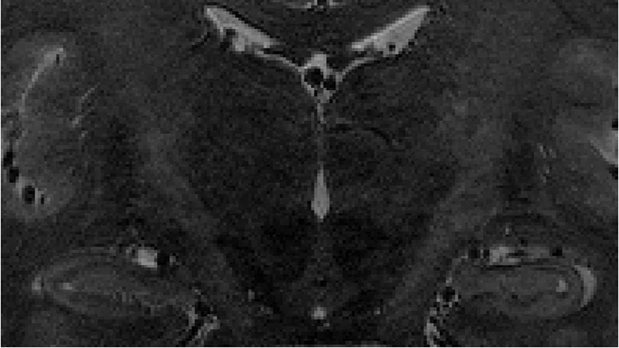
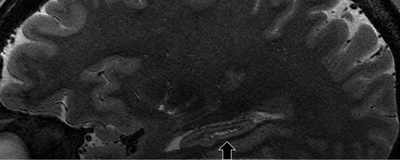
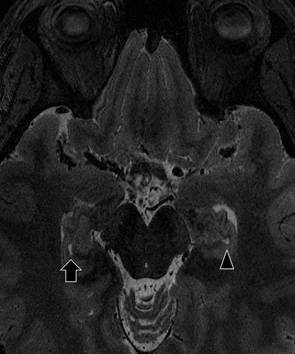
Results: Both observers identified HSRs with 7T MR images in all subjects. Excellent interobserver agreement was shown (κ = 1.0). The shape of HSRs was variable (spot-like, curvilinear, ovoid, or beaded appearance). Volumes of the HSRs were not correlated with age.

Conclusions: Hippocampal sulcus remnants are commonly seen in healthy nonelderly adults using 7T MR imaging. Accurate diagnosis of HSR based on the microanatomy of hippocampus makes it easier to differentiate them from lesions, and it may help prevent unnecessary treatment.


Conflict of interest statement

Disclosure of interest

The authors declare that there is no relationships/conditions/circumstances that present a potential conflict of interest in this manuscript.

Figures

Fig. 1
Fig. 1
Example of the voxel of interests (VOIs). Source image (a). The VOIs are indicated colored blocks (b).
Fig. 1
Fig. 1
Example of the voxel of interests (VOIs). Source image (a). The VOIs are indicated colored blocks (b).
Fig. 2
Fig. 2
21-year-old male. Sagittal (a), coronal (b), and transverse planes (c). The hippocampal sulcus remnants (HSRs) are identified in the hippocampal tail.
Fig. 2
Fig. 2
21-year-old male. Sagittal (a), coronal (b), and transverse planes (c). The hippocampal sulcus remnants (HSRs) are identified in the hippocampal tail.
Fig. 2
Fig. 2
21-year-old male. Sagittal (a), coronal (b), and transverse planes (c). The hippocampal sulcus remnants (HSRs) are identified in the hippocampal tail.
Fig. 3
Fig. 3
26-year-old female. Sagittal (a) and transverse planes (b). The shape of the right HSRs (arrow) appear to be beaded structure on the sagittal image. The left HSR (arrowhead) is also identified on the transverse image.
Fig. 3
Fig. 3
26-year-old female. Sagittal (a) and transverse planes (b). The shape of the right HSRs (arrow) appear to be beaded structure on the sagittal image. The left HSR (arrowhead) is also identified on the transverse image.
Fig. 4
Fig. 4
51-year-old male. Magnified coronal (a) and continuous sagittal images (b-d). The sagittal images indicate the tubular hypointense structure (arrow) within the HSR (arrowhead), which seems to be blood vessel.
Fig. 4
Fig. 4
51-year-old male. Magnified coronal (a) and continuous sagittal images (b-d). The sagittal images indicate the tubular hypointense structure (arrow) within the HSR (arrowhead), which seems to be blood vessel.
Fig. 4
Fig. 4
51-year-old male. Magnified coronal (a) and continuous sagittal images (b-d). The sagittal images indicate the tubular hypointense structure (arrow) within the HSR (arrowhead), which seems to be blood vessel.
Fig. 4
Fig. 4
51-year-old male. Magnified coronal (a) and continuous sagittal images (b-d). The sagittal images indicate the tubular hypointense structure (arrow) within the HSR (arrowhead), which seems to be blood vessel.
Fig. 5
Fig. 5
Scatter plots of the age and the volume of the HSR. Note that the volume of the HSR is not correlated with age (r = 0.02, p = 0.88)
Fig. 6
Fig. 6
Scatter plots of the right and the left HSR volumes. Solid and dashed lines represent linear regression of the plots and 95% confidence intervals, respectively. There is the significant correlation between the volume of the HSR bilaterally (r = 0.62, p = 0.0001).

References

    1. Humphrey T The development of the human hippocampal fissure. J Anat 1967;101:655–676 - PMC - PubMed
    1. Barboriak DP, Doraiswamy PM, Krishnan KR, et al. Hippocampal sulcal cavities on MRI: relationship to age and apolipoprotein E genotype. Neurology 2000;54:2150–2153 - PubMed
    1. Dekeyzer S, De Kock I, Nikoubashman O, et al. “Unforgettable” - a pictorial essay on anatomy and pathology of the hippocampus. Insights Imaging 2017;8:199–212 - PMC - PubMed
    1. Erdem A, Yasargil G, Roth P. Microsurgical anatomy of the hippocampal arteries. Journal of neurosurgery 1993;79:256–265 - PubMed
    1. Sasaki M, Sone M, Ehara S, et al. Hippocampal sulcus remnant: potential cause of change in signal intensity in the hippocampus. Radiology 1993;188:743–746 - PubMed