. 2019 Dec 2;14(12):e0224921.
doi: 10.1371/journal.pone.0224921. eCollection 2019.

Three-dimensional analysis of pancreatic fat by fat-water magnetic resonance imaging provides detailed characterization of pancreatic steatosis with improved reproducibility

Affiliations

Three-dimensional analysis of pancreatic fat by fat-water magnetic resonance imaging provides detailed characterization of pancreatic steatosis with improved reproducibility

Shingo Kato et al. PLoS One. .

Abstract

Background: Since pancreatic steatosis is reported as a possible risk factor for pancreatic cancer, the development of a non-invasive method to quantify pancreatic steatosis is needed. Proton density fat fraction (PDFF) measurement is a magnetic resonance imaging (MRI) based method for quantitatively assessing the steatosis of a region of interest (ROI). Although it is commonly used for quantification of hepatic steatosis, pancreatic PDFF can greatly vary depending on the ROI's location because of the patchy nature of pancreatic fat accumulation. In this study, we attempted to quantify pancreatic steatosis by fat-water MRI with improved reproducibility.

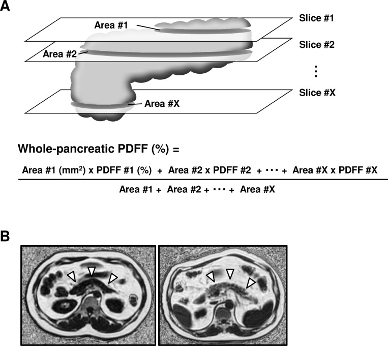
Methods: Using the MRI images of 159 patients with nonalcoholic fatty liver disease, we attempted to calculate the average PDFF of whole pancreas. We set ROIs covering the entire area of the pancreas appearing in every slice and calculated the average PDFF from all the voxels included in the pancreas. We named this average value as whole-pancreatic PDFF and evaluated the reproducibility of the measured values. In addition to whole-pancreatic PDFF, we measured the average PDFF of the pancreatic head (head-PDFF) and that of the pancreatic body plus tail separately and analyzed their correlation with the clinical characteristics of the patients.

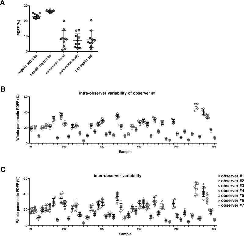
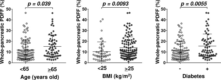
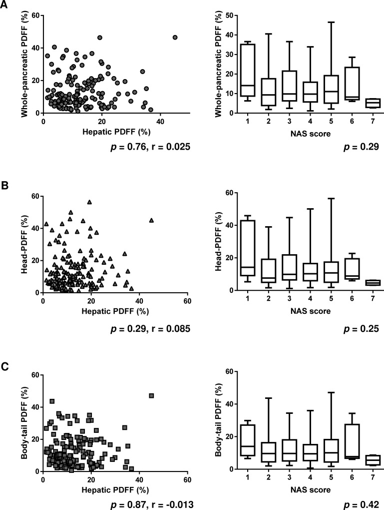
Results: The mean inter-examiner coefficient of variation of the whole-pancreatic PDFF was 11.39%. The whole-pancreatic PDFF was correlated with age (p = 0.039), body mass index (p = 0.0093) and presence/absence of diabetes (p = 0.0055). The serum level of low-density lipoprotein cholesterol was inversely correlated with the head-PDFF.

Conclusion: We developed a new measurement method of the pancreatic PDFF with greater reproducibility. Using this method, we characterized pancreatic steatosis in detail. This novel measurement method allows accurate estimation of the severity of pancreatic steatosis and is therefore useful for the detailed characterization of pancreatic steatosis.


Conflict of interest statement

The authors have declared that no competing interests exist.

Figures

Fig 1
Fig 1. The schema of whole-pancreatic analysis and representative fat-water MRI images of low or high degree of pancreatic steatosis.
(A) The schema of whole-pancreatic analysis is shown. (B) Representative pancreatic images of fat-water MRI are shown. A white arrow indicates the pancreas. The left panel shows the low degree of pancreatic steatosis in a sample whose whole-pancreatic FF value was determined to be 1.20%. The right panel shows the high degree of pancreatic steatosis in a sample whose whole-pancreatic FF value was determined to be 34.87%.
Fig 2
Fig 2. Evaluation of reproducibility of each measurement method.
(A) The representative result of the evaluation of the variations in values of hepatic or pancreatic PDFF depending on the positions of the ROIs is shown. The values of the pancreatic PDFF measured using small ROIs varied widely depending on the locations of the ROIs. (B) All measurements by observer #1 for intra-observer variability analysis are shown. (C) All measurements by 6 observers for inter-observer variability analysis are shown. The average value of five repeated measurements by each observer was taken as the measured value of the observer.
Fig 3
Fig 3. Correlation between whole-pancreatic PDFF and the clinical characteristics.
Whole-pancreatic FF was correlated with age (left panel, p = 0.039), BMI (middle panel, p = 0.0093), and presence/absence of diabetes (right panel, p = 0.0055).
Fig 4
Fig 4. Correlation between whole-pancreatic PDFF and the hepatic PDFF or NAS score.
Whole-pancreatic PDFF was not correlated with hepatic PDFF (left panel, p = 0.76), and NAS score (right panel, p = 0.29). (B) Head-PDFF was not correlated with hepatic PDFF (left panel, p = 0.29), and NAS score (right panel, p = 0.25). (C) Body-tail PDFF was not correlated with hepatic PDFF (left panel, p = 0.87), and NAS score (right panel, p = 0.42).

References

    1. Takahashi M, Hori M, Ishigamori R, Mutoh M, Imai T, Nakagama H. Fatty pancreas: A possible risk factor for pancreatic cancer in animals and humans. Cancer Sci. 2018;109(10):3013–23. 10.1111/cas.13766 - DOI - PMC - PubMed
    1. Hori M, Takahashi M, Hiraoka N, Yamaji T, Mutoh M, Ishigamori R, et al. Association of pancreatic Fatty infiltration with pancreatic ductal adenocarcinoma. Clinical and translational gastroenterology. 2014;5:e53 10.1038/ctg.2014.5 - DOI - PMC - PubMed
    1. Olsen TS. Lipomatosis of the pancreas in autopsy material and its relation to age and overweight. Acta Pathol Microbiol Scand A. 1978;86A(5):367–73. 10.1111/j.1699-0463.1978.tb02058.x . - DOI - PubMed
    1. Stamm BH. Incidence and diagnostic significance of minor pathologic changes in the adult pancreas at autopsy: a systematic study of 112 autopsies in patients without known pancreatic disease. Human pathology. 1984;15(7):677–83. 10.1016/s0046-8177(84)80294-4 . - DOI - PubMed
    1. Hu HH, Kim HW, Nayak KS, Goran MI. Comparison of fat-water MRI and single-voxel MRS in the assessment of hepatic and pancreatic fat fractions in humans. Obesity. 2010;18(4):841–7. 10.1038/oby.2009.352 - DOI - PMC - PubMed