. 2019 Dec 2;14(12):e0225767.
doi: 10.1371/journal.pone.0225767. eCollection 2019.

Respiratory syncytial virus exhibits differential tropism for distinct human placental cell types with Hofbauer cells acting as a permissive reservoir for infection

Affiliations

Respiratory syncytial virus exhibits differential tropism for distinct human placental cell types with Hofbauer cells acting as a permissive reservoir for infection

Vladimir Bokun et al. PLoS One. .

Abstract

Background: Respiratory syncytial virus (RSV) is capable of transient viremia and extrapulmonary dissemination. Recently, this virus has been identified in fetal cord blood, suggesting the possibility of in utero acquisition in humans. Here, we assess permissivity and kinetics of RSV replication in primary human placental cells, examine their potential to transfer this infection to neighboring cells, and measure the inflammatory response evoked by the virus.

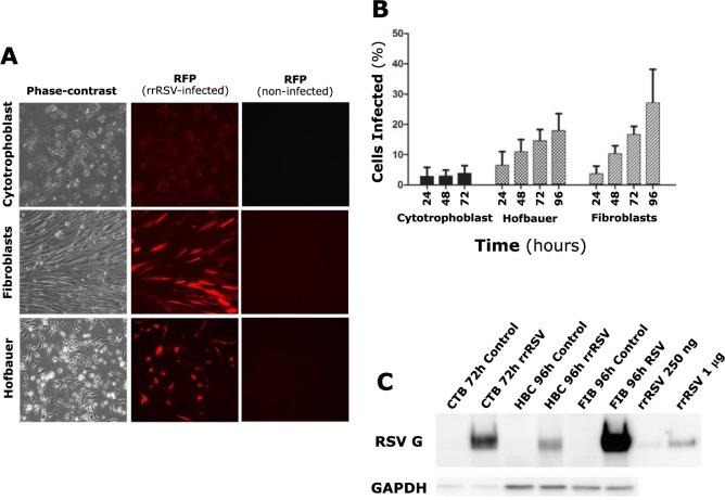
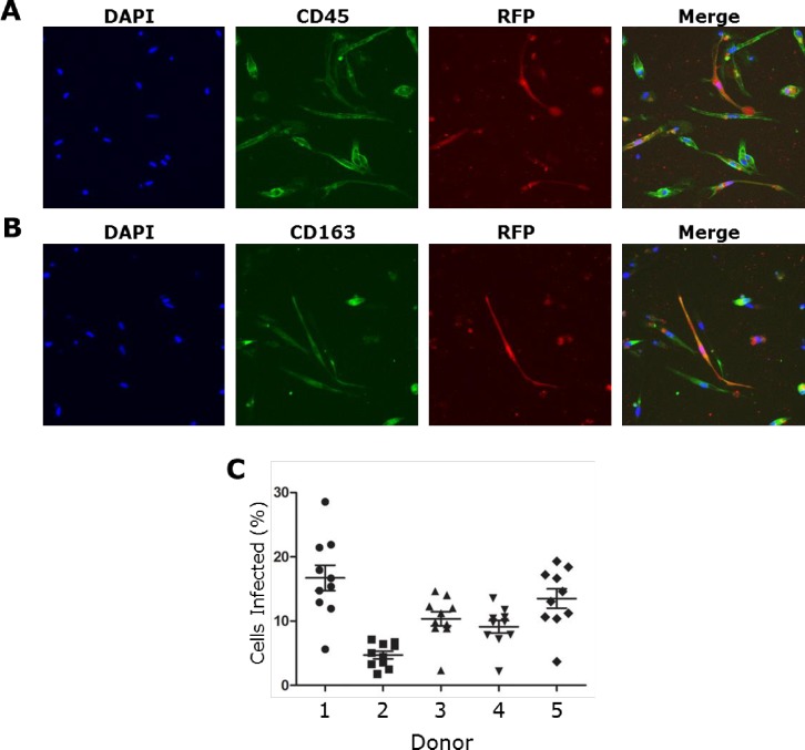
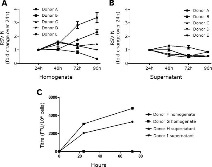
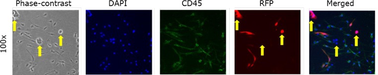
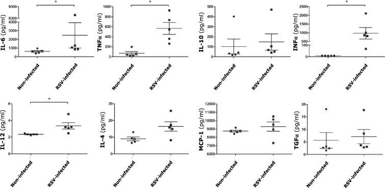
Methods and findings: Human placental villus tissue was collected immediately upon delivery and processed for isolation of placental cytotrophoblast, fibroblast, and macrophage (Hofbauer) cells. Isolated cells were infected with a recombinant RSV-A2 strain (rrRSV) expressing red fluorescent protein (RFP) and analyzed by fluorescence microscopy, Western blot, and quantitative PCR (qPCR). Based on RFP expression, rrRSV exhibited differential tropism for the three major placental cell types. Placental fibroblasts and Hofbauer cells were permissive and supported productive rrRSV replication. While infected cytotrophoblast cells expressed viral glycoprotein (G protein), only limited RSV replication was detected. Importantly, qPCR and fluorescence-focused unit assay revealed that the viral progeny remains trapped within infected Hofbauer cells for up to 30 days, with no release into surrounding media. Yet, Hofbauer cells passed the infection onto overlaid naïve 16HBE cells, suggesting contact-dependent trans-infection. Lastly, a significant increase in proinflammatory cytokines, particularly IL-6, TNF-alpha, and IFN-gamma was measured in the supernatant of infected Hofbauer cells by multiplex cytokine assay and conventional ELISA.

Conclusions: This study demonstrates that RSV can replicate in human placenta, exhibits differential tropism for distinct placental cell types, can be stored and transferred to neighboring cells by Hofbauer cells, and elicits an inflammatory response. It also supports the hypothesis that this respiratory virus can be vertically transferred to the fetus and potentially affect its development and the outcome of pregnancies.


Conflict of interest statement

The authors have declared that no competing interests exist.

Figures

Fig 1
Fig 1. RSV infection of primary human placental cells.
(A) Isolated human placental cells (left panels) after 72 hours (cytotrophoblast cells) or 96 hours (fibroblast and Hofbauer cells) infection at MOI of 1.0 with an RSV-A2 strain (rrRSV) expressing red fluorescent protein (RFP). Strong RFP expression resulting from rrRSV replication was observed in fibroblast and Hofbauer, but not in cytotrophoblast cells (center panels). Matched non-infected controls did not exhibit any fluorescence (right panels). (B) Proportion of cytotrophoblast, fibroblast, and Hofbauer cells exhibiting red fluorescence as a result of rrRSV replication. Each time-point represents the mean ± SEM of 8 different fields. (C) Expression of RSV G protein in cytotrophoblast (CTB), Hofbauer (HBC), and fibroblast (FIB) cells infected with rrRSV and processed at 72 or 96 hours for Western blot analysis. rrRSV lysate was used in two different concentrations (250 ng and 1 μg) as a positive control.
Fig 2
Fig 2. RSV infection of Hofbauer cells.
Hofbauer cells were infected with rrRSV at MOI of 1 for 96 hours and then fixed with 4% paraformaldehyde. RFP expression by replicating rrRSV co-localized with (A) myeloid cell marker CD45 and (B) macrophage cell marker CD163 in infected Hofbauer cells. (C) Proportion of Hofbauer cells from 5 different donors exhibiting red fluorescence as a result of rrRSV replication. Infectivity was variable among donors and ranged from approximately 5 to 17 percent. Data are shown as mean ± SEM of 5 different donors.
Fig 3
Fig 3. RSV infection kinetics in Hofbauer cells.
(A) Expression of RSV nucleocapsid (N) RNA transcripts measured in lysates of Hofbauer cells infected with rrRSV at MOI of 1 using ß-actin as housekeeping gene. Fold changes were calculated using the 24-hour time-point as baseline to demonstrate growth kinetics between cell homogenate and supernatants. Data are shown as mean ± SEM of 5 different donors. (B) RSV N RNA transcripts in supernatants matching the cell homogenates shown in panel A and normalized based on input volume. RSV N transcription increased in cell homogenates of 4 of the 5 analyzed donors, whereas no increase was observed in the corresponding supernatants. (C) Viral titers measured at 24- and 72-hours post-infection with rrRSV at MOI of 1 using a fluorescence-focused unit (FFU) assay. Whereas viral replication within Hofbauer cells was detected at 24 hours and increased by 72 hours, no virus was found in the corresponding supernatants at either time-point.
Fig 4
Fig 4. Direct transmission of RSV from Hofbauer cells to naïve 16HBE.
Hofbauer cells were infected at a MOI of 0.01 and incubated for 48 hours, after which uninfected 16HBE cells were seeded for co-culture. After 72 hours of co-culture, the cells were fixed for analysis of RFP expression by fluorescence microscopy. The characteristic epithelial “colonies” formed by 16HBE cells (yellow arrows) and the CD45 staining (green) identifying Hofbauer cells allowed to recognize the two cell types. RFP expression (red) and syncytia formation was observed in 16HBE cells at 72 hours, indicating transmission of the infection from the Hofbauer cells to the epithelial cells.
Fig 5
Fig 5. Cytokine expression of Hofbauer cells following infection with RSV.
Cytokines concentration in supernatants collected at 24 hours post-infection with rrRSV at MOI of 1 were measured using the Luminex/Magpix magnetic bead platform, with the exception of TNF-alpha that was measured by conventional sandwich ELISA. The supernatants of infected Hofbauer cells exhibited significantly increased expression of the proinflammatory cytokines IL-6, TNF-alpha, IFN-gamma, and IL-12. Data are mean ± SEM; n = 5 donors; *p<0.05 vs. non-infected controls).

References

    1. Campbell AP, Chien JW, Kuypers J, Englund JA, Wald A, Guthrie KA, et al. Respiratory virus pneumonia after hematopoietic cell transplantation (HCT): associations between viral load in bronchoalveolar lavage samples, viral RNA detection in serum samples, and clinical outcomes of HCT. J Infect Dis. 2010;201:1404–13. 10.1086/651662 - DOI - PMC - PubMed
    1. Halfhide CP, Flanagan BF, Brearey SP, Hunt JA, Fonceca AM, McNamara PS, et al. Respiratory syncytial virus binds and undergoes transcription in neutrophils from the blood and airways of infants with severe bronchiolitis. J Infect Dis. 2011;204:451–8. 10.1093/infdis/jir280 - DOI - PMC - PubMed
    1. O'Donnell DR, McGarvey MJ, Tully JM, Balfour-Lynn IM, Openshaw PJ. Respiratory syncytial virus RNA in cells from the peripheral blood during acute infection. J Pediatr. 1998;133:272–4. 10.1016/s0022-3476(98)70234-3 - DOI - PubMed
    1. Rezaee F, Gibson LF, Piktel D, Othumpangat S, Piedimonte G. Respiratory syncytial virus infection in human bone marrow stromal cells. Am J Respir Cell Mol Biol. 2011;45:277–86. 10.1165/rcmb.2010-0121OC - DOI - PMC - PubMed
    1. Waghmare A, Campbell AP, Xie H, Seo S, Kuypers J, Leisenring W, et al. Respiratory syncytial virus lower respiratory disease in hematopoietic cell transplant recipients: viral RNA detection in blood, antiviral treatment, and clinical outcomes. Clin Infect Dis. 2013;57:1731–41. 10.1093/cid/cit639 - DOI - PMC - PubMed

Publication types