Review
. 2020 Jan:29:101394.
doi: 10.1016/j.redox.2019.101394. Epub 2019 Nov 26.

Teaching the basics of the mechanism of doxorubicin-induced cardiotoxicity: Have we been barking up the wrong tree?

Affiliations
Review

Teaching the basics of the mechanism of doxorubicin-induced cardiotoxicity: Have we been barking up the wrong tree?

Balaraman Kalyanaraman. Redox Biol. 2020 Jan.

Abstract

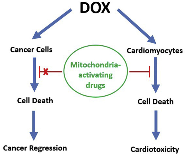
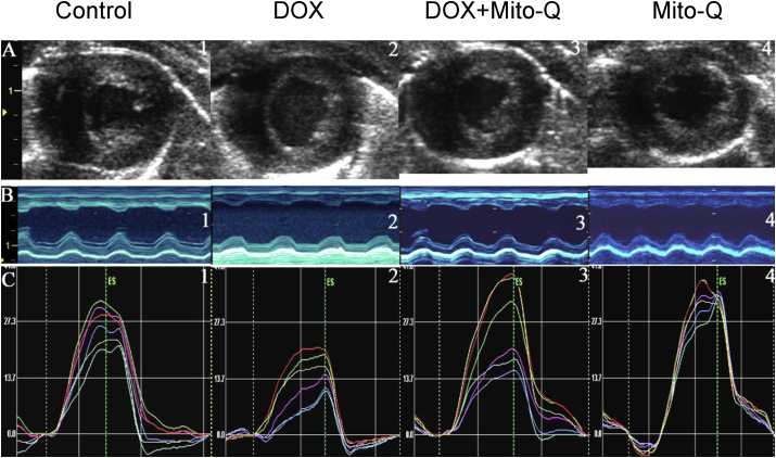
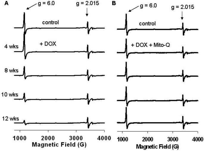
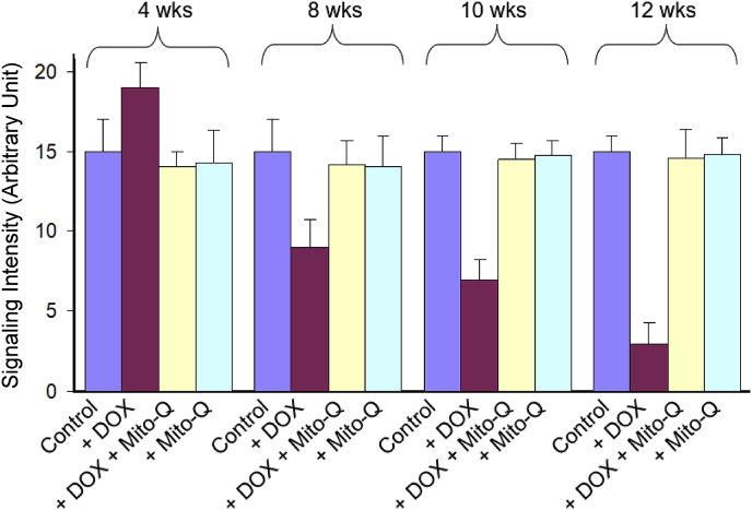
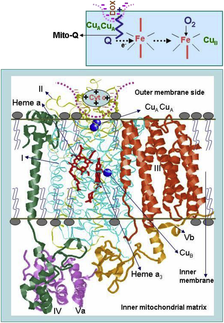
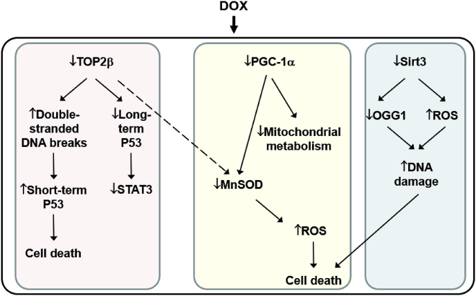
Doxorubicin (DOX), or Adriamycin, an anthracycline antibiotic discovered serendipitously as a chemotherapeutic drug several decades ago, is still one of the most effective drugs for treating various adult and pediatric cancers (breast cancer, Hodgkin's disease, lymphoblastic leukemia). However, one of the major side effects of the continuous use of DOX is dose-dependent, long-term, and potentially lethal cardiovascular toxicity (congestive heart failure and cardiomyopathy) in cancer survivors many years after cessation of chemotherapy. In addition, predisposition to cardiotoxicity varied considerably among individuals. The long-held notion that DOX cardiotoxicity is caused by reactive oxygen species formed from the redox-cycling of DOX semiquinone lacks rigorous proof in a chronic animal model, and administration of reactive oxygen species detoxifying agents failed to reverse DOX-induced cardiac problems. In this review, I discuss the pros and cons of the reactive oxygen species pathway as a primary or secondary mechanism of DOX cardiotoxicity, the role of topoisomerases, and the potential use of mitochondrial-biogenesis-enhancing compounds in reversing DOX-induced cardiomyopathy. New approaches for well-designed clinical trials that repurpose FDA-approved drugs and naturally occurring polyphenolic compounds prophylactically to prevent or mitigate cardiovascular complications in both pediatric and adult cancer survivors are needed. Essentially, the focus should be on enhancing mitochondrial biogenesis to prevent or mitigate DOX-induced cardiotoxicity.

Keywords: Cardio-oncology; Cardioprotection; Chemotherapy; Mitochondrial biogenesis; Reactive oxygen species; Topoisomerase.


Conflict of interest statement

The authors declare that they have no known competing financial interests or personal relationships that could have appeared to influence the work reported in this paper.

Figures

Image 1
Graphical abstract
Fig. 1
Fig. 1
Chemical structure of DOX/Adriamycin. The functional groups that are thought to be critical to the DOX mechanism of action are marked.
Fig. 2
Fig. 2
Redox-cycling of DOX semiquinone and ROS-induced mechanism of mitochondrial oxidation and cardiotoxicity. Reprinted by permission from Springer Nature Customer Service Centre GmbH: Springer Nature Molecular and Cellular Biochemistry (Doxorubicin-induced apoptosis: Implications in cardiotoxicity Kalyanaraman B, Joseph J, Kalivendi S, Wang S, Konorev E, Kotamraju S), Kluwer Academic Publishers (2002).
Fig. 3
Fig. 3
DOX-induced cardiomyopathy in a rat model and the cardioprotective effect of Mito-Q. Echocardiography, EPR, and biochemical measurements were performed weekly. Reprinted from Biophysics Journal, 96, Chandran K, Aggarwal D, Migrino RQ, Joseph J, McAllister D, Konorev EA, Antholine WE, Zielonka J, Srinivasan S, Avadhani NG, Kalyanaraman B, Doxorubicin inactivates myocardial cytochrome c oxidase in rats: Cardioprotection by Mito-Q, 1388–1398, Biophysical Society (2009), with permission from Elsevier.
Fig. 4
Fig. 4
Mito-Q inhibits DOX-induced cardiac dysfunction—two-dimensional strain echocardiography measurements. (A) End systolic two-dimensional B-mode images at mid- ventricular level. (B) Anatomical M-mode through the anterior and interior walls. (C) The radial strain from six equidistant segments of the left ventricle. Reprinted from Ultrasound in Medicine & Biology, 34, Migrino RQ, Aggarwal D, Konorev E, Brahmbhatt T, Bright M, Kalyanaraman B, Early detection of doxorubicin cardiomyopathy using two-dimensional strain echocardiography, 208–214, World Federation for Ultrasound in Medicine & Biology (2008), with permission from Elsevier.
Fig. 5
Fig. 5
Time-course EPR spectra of myocardial tissues. Low-temperature EPR of heart tissues isolated from rats treated with (A) DOX and (B) DOX and Mito-Q. Reprinted from Biophysics Journal, 96, Chandran K, Aggarwal D, Migrino RQ, Joseph J, McAllister D, Konorev EA, Antholine WE, Zielonka J, Srinivasan S, Avadhani NG, Kalyanaraman B, Doxorubicin inactivates myocardial cytochrome c oxidase in rats: Cardioprotection by Mito-Q, 1388–1398, Biophysical Society (2009), with permission from Elsevier.
Fig. 6
Fig. 6
Time-course EPR spectral intensity (by monitoring the signal at g = 6.0). Reprinted from Biophysics Journal, 96, Chandran K, Aggarwal D, Migrino RQ, Joseph J, McAllister D, Konorev EA, Antholine WE, Zielonka J, Srinivasan S, Avadhani NG, Kalyanaraman B, Doxorubicin inactivates myocardial cytochrome c oxidase in rats: Cardioprotection by Mito-Q, 1388–1398, Biophysical Society (2009), with permission from Elsevier.
Fig. 7
Fig. 7
DOX-induced inactivation of cytochrome c oxidase:proposed model. Reprinted from Biophysics Journal, 96, Chandran K, Aggarwal D, Migrino RQ, Joseph J, McAllister D, Konorev EA, Antholine WE, Zielonka J, Srinivasan S, Avadhani NG, Kalyanaraman B, Doxorubicin inactivates myocardial cytochrome c oxidase in rats: Cardioprotection by Mito-Q, 1388–1398, Biophysical Society (2009), with permission from Elsevier.
Fig. 8
Fig. 8
Lack of effect of Mito-Q on oxidative inactivation of aconitase. Reprinted from Biophysics Journal, 96, Chandran K, Aggarwal D, Migrino RQ, Joseph J, McAllister D, Konorev EA, Antholine WE, Zielonka J, Srinivasan S, Avadhani NG, Kalyanaraman B, Doxorubicin inactivates myocardial cytochrome c oxidase in rats: Cardioprotection by Mito-Q, 1388–1398, Biophysical Society (2009), with permission from Elsevier.
Fig. 9
Fig. 9
The current mechanistic model for DOX-induced cardiotoxicity.

References

    1. Rosen M.R., Myerburg R.J., Francis D.P., Cole G.D., Marban E. Translating stem cell research to cardiac disease therapies: pitfalls and prospects for improvement. J. Am. Coll. Cardiol. 2014;64:922–937. - PMC - PubMed
    1. Sheng C.C., Amiri-Kordestani L., Palmby T., Force T., Hong C.C., Wu J.C., Croce K., Kim G., Moslehi J. 21st century cardio-oncology: identifying cardiac safety signals in the era of personalized medicine. JACC Basic Transl Sci. 2016;1:386–398. - PMC - PubMed
    1. Hardaway B.W. Adriamycin-associated cardiomyopathy: where are we now? updates in pathophysiology, dose recommendations, prognosis, and outcomes. Curr. Opin. Cardiol. 2019;34:289–295. - PubMed
    1. Young R.C., Ozols R.F., Myers C.E. The anthracycline antineoplastic drugs. N. Engl. J. Med. 1981;305:139–153. - PubMed
    1. Blum R.H., Carter S.K. Adriamycin. A new anticancer drug with significant clinical activity. Ann. Intern. Med. 1974;80:249–259. - PubMed

Publication types