Review
. 2020 Feb:60:17-26.
doi: 10.1016/j.sbi.2019.09.007. Epub 2019 Nov 29.

Molecular structure in biomolecular condensates

Affiliations
Review

Molecular structure in biomolecular condensates

Ivan Peran et al. Curr Opin Struct Biol. 2020 Feb.

Abstract

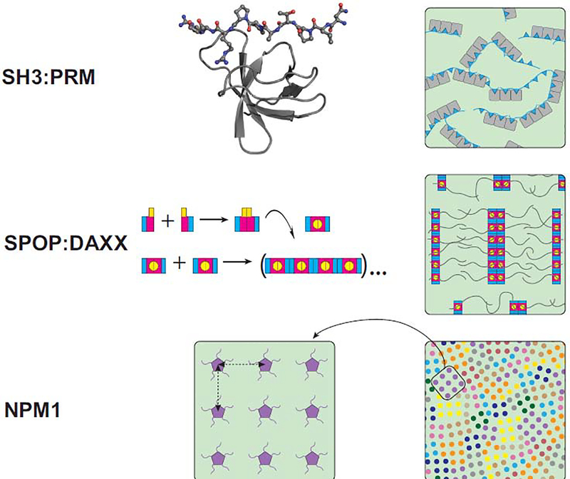
Evidence accumulated over the past decade provides support for liquid-liquid phase separation as the mechanism underlying the formation of biomolecular condensates, which include not only 'membraneless' organelles such as nucleoli and RNA granules, but additional assemblies involved in transcription, translation and signaling. Understanding the molecular mechanisms of condensate function requires knowledge of the structures of their constituents. Current knowledge suggests that structures formed via multivalent domain-motif interactions remain largely unchanged within condensates. Two different viewpoints exist regarding structures of disordered low-complexity domains within condensates; one argues that low-complexity domains remain largely disordered in condensates and their multivalency is encoded in short motifs called 'stickers', while the other argues that the sequences form cross-β structures resembling amyloid fibrils. We review these viewpoints and highlight outstanding questions that will inform structure-function relationships for biomolecular condensates.


Figures

Figure 1.
Figure 1.. Domain architecture determines super-molecular structure in dense phases.
(Top row) Insight into dense phase structures via high-resolution structures of domain/motif interactions. (Left) A high resolution crystal structure of an SH3 domain bound to a proline-rich motif (PRM) peptide. (PDB: 4WCI) [75]. (Right) A cartoon representation of a dense phase (green background) generated by multivalent SH3:PRM interactions. Three SH3 domains are connected by linkers in a single molecule (gray); a binding partner containing three proline-rich motifs (cyan) interacts with the SH3-protein via multivalent interactions. The two protein types crosslink each other. (Middle row) Multivalency via linear polymerization. (Left) SPOP monomers dimerize and the dimers polymerize into linear structures. The BACK dimerization domain is shown in cyan, the BTB dimerization domain in magenta, and the MATH substrate binding domain in yellow. The curved arrow indicates a 90° rotation of a SPOP dimer onto its side. (Right) SPOP oligomers bind several DAXX molecules via their MATH domains, giving rise to “brush-like” structures. These brushes are crosslinked via DAXX interactions [16,30]. (Bottom row) Local order in a liquid dense phase. The distances between NPM1 pentamers (magenta) repeat (left), giving rise to locally ordered arrays of Npm1 molecules within the dense phase. Npm1 molecules are crosslinked by arginine-rich peptides (not shown). (Right) Over greater distances, the anisotropic arrangement of arrays (each shown in different colors) gives rise to global disorder and liquid-like behavior [28].
Figure 2.
Figure 2.. LCDs can form different types of assemblies.
(a) LLPS of intrinsically disordered LCDs. From left to right: (1) A sample with a large dense drop at the bottom of the tube (dark green) overlaid by the dilute phase (light green) is prepared from (2) many small micron-sized droplets (dark green) that fuse. (3) The protein chains remain largely disordered in the dense phase (green mesh). Interactions driving phase separation include hydrophobic, π-π interactions (e.g. between aromatic residues shown in magenta stick representation), cation-π interactions between aromatics (magenta sticks) and positively charged residues (blue sticks), polar interactions (violet sticks), electrostatic interactions between positive (blue sticks) and negative (red sticks) charges, and hydrogen bonding (orange sticks). (b) Hydrogels of LCDs. From left to right: (1) Many low complexity domains such as that of FUS can assemble into hydrogels. (2) The FUS hydrogel is composed of amyloid-like fibrils. (3) Solid-state NMR structure of the core region of FUS that assembles into long filaments with a cross-β architecture. The remaining 77% of the LCD sequence remains disordered, indicated as red lines. (4) The atomic structure of a FUS monomer within a fibril reveals short in-register β-sheets separated by loops. Of note is the absence of all but one hydrophobic residue (yellow). (c) Irreversible amyloid fibrils formed by typical amyloidogenic proteins, not LCDs. (1) Cartoon depiction of irreversible fibrils sedimented in an Eppendorf tube. (2) TEM of α-synuclein fibrils. (3) Structure of the components of the fibrils indicates a long, continuous β-sheet core flanked by disordered regions. (4) Atomic structure of the core of an α-synuclein monomer within a fibril shows an in-register β-sheet stabilized by hydrophobic contacts involving Val, Ile, Ala and Phe residues (yellow). (Some illustrations were made in BioRender (biorender.com).)
Figure 3:
Figure 3:. (Super-)molecular structural properties that define dense phases.
Clockwise from the top: (1) What are the conformations adopted in phase separation-mediating contacts and what is the rearrangement of building blocks relative to each other (also see Figure 1)? (2) Do the stabilities of folded protein domains differ between the dilute and dense phase? And are the affinities between domains and motifs different between the phases? (3) What is the extent of ordering inside of droplets? (4) What are the lifetimes of individual interactions within the dense phase? (5) Over what distance within the dense phase is the movement of building blocks correlated? (6) How many cross-links form between molecules in the dense phase and what is the resulting mesh size? (7) Do the structures of molecules and their assemblies differ between the bulk dense phase and the phase boundary with the dilute phase?

References

    1. Case LB, Zhang X, Ditlev JA, Rosen MK: Stoichiometry controls activity of phase-separated clusters of actin signaling proteins. Science 2019, 363:1093–1097. - PMC - PubMed
    1. Ries RJ, Zaccara S, Klein P, Olarerin-George A, Namkoong S, Pickering BF, Patil DP, Kwak H,Lee JH, Jaffrey SR: m(6)A enhances the phase separation potential of mRNA. Nature 2019, 571:424–428. - PMC - PubMed
    1. Franzmann TM, Jahnel M, Pozniakovsky A, Mahamid J, Holehouse AS, Nuske E, Richter D, Baumeister W, Grill SW, Pappu RV, et al. : Phase separation of a yeast prion protein promotes cellular fitness. Science 2018, 359. - PubMed
    1. Boija A, Klein IA, Sabari BR, Dall’Agnese A, Coffey EL, Zamudio AV, Li CH, Shrinivas K, Manteiga JC, Hannett NM, et al. : Transcription Factors Activate Genes through the Phase-Separation Capacity of Their Activation Domains. Cell 2018, 175:1842–1855 e1816. - PMC - PubMed
    1. Brangwynne CP, Eckmann CR, Courson DS, Rybarska A, Hoege C, Gharakhani J, Julicher F, Hyman AA: Germline P granules are liquid droplets that localize by controlled dissolution/condensation. Science 2009, 324:1729–1732. - PubMed

Publication types

MeSH terms

Substances

LinkOut - more resources