. 2019 Dec 2;19(1):266.
doi: 10.1186/s12903-019-0970-7.

Immediate implant placement in anterior teeth with grafting material of autogenous tooth bone vs xenogenic bone

Affiliations

Immediate implant placement in anterior teeth with grafting material of autogenous tooth bone vs xenogenic bone

Dong Wu et al. BMC Oral Health. .

Abstract

Background: The aim of the study was to compare the efficacy of the autogenous tooth bone and xenogenic bone grafted in immediate implant placement with bone defect.

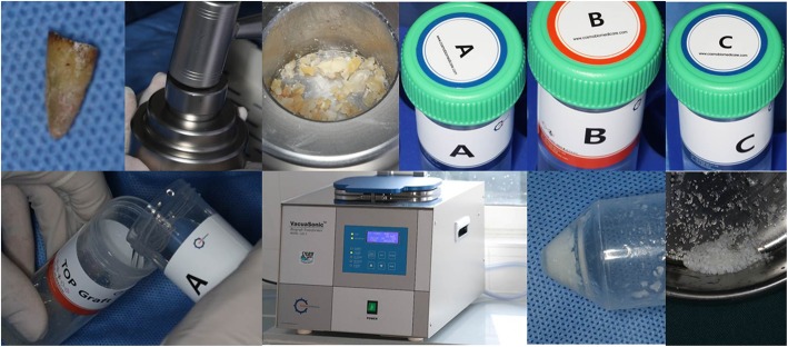
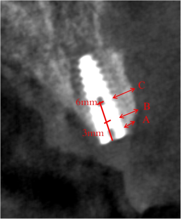
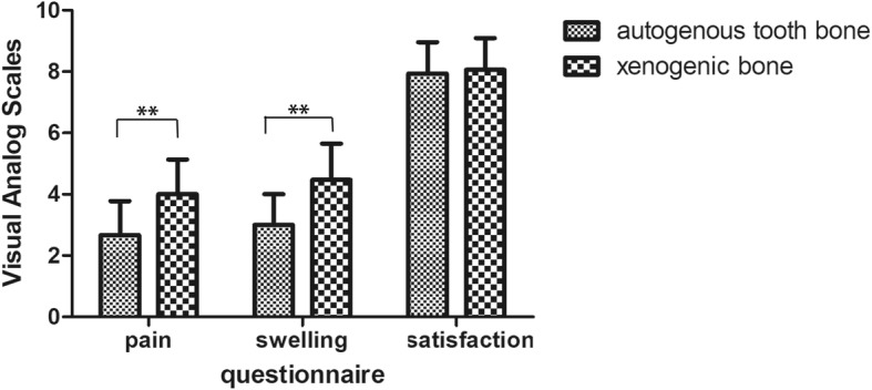
Methods: Thirty patients whose compromised anterior teeth need immediate implant placement were enrolled. Autogenous tooth bone made from the extracted teeth by chair-side or the xenogenic bone were used to repaired bone defect. Clinical examination, radiographic assessment about the horizontal bone change in the level of 0 mm, 3 mm and 6 mm below the implant neck and the marginal bone loss were made immediately, 6 and 12 months after implant placement. Questionnaire of the feelings about the surgery were made at the time of removing the sutures.

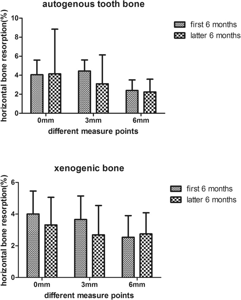
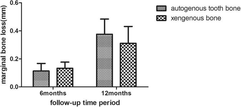
Results: All implants achieved the success criteria without any complications at the follow-up period. The percent of the horizontal bone change and the marginal bone loss at 6 and 12 months were almost the same between two groups (P > .05). The horizontal bone loss at the first or the latter 6 months was almost the same (P > .05). But the horizontal bone loss at the 6 mm level was less than the 0 mm and 3 mm levels at 6 and 12 months (P < .05). Meanwhile patients seem more satisfied with the autogenous tooth bone derived from the questionnaire.

Conclusion: The bone volume change in the facial part of the implant after immediate placement is almost the same between two groups. Providing clinical evidence that the autogenous tooth bone made from compromised tooth can be an acceptable bone graft material.

Keywords: Autogenous tooth; Bone graft; Immediate implant placement; Implant dentistry.


Conflict of interest statement

The authors declare that they have no competing interests.

Figures

Fig. 1
Fig. 1
Chairside preparation of autogenous tooth bone graft materials. Autogenous tooth without retention value was removed with minimally invasive extraction. Surface preparation and pretreatment were performed on the teeth. The tooth was crushed into debris. Bone powder was prepared. Demineralization, sterilization and rinse were performed for autogenous tooth bone graft materials
Fig. 2
Fig. 2
Surgical proceduce of the implant placement and bone grafting
Fig. 3
Fig. 3
The measured point of facial bone. a represent the bone width at the level of implant neck or the top of the buccal bone. b and c represent the bone width of the 3 mm and 6 mm below the implant neck level
Fig. 4
Fig. 4
The change of the horizontal bone at 6 and 12 months in the level of 0 mm, 3 mm and 6 mm between the autogenous tooth bone and xenogenic bone
Fig. 5
Fig. 5
The change of the horizontal bone at the group of autogenous tooth bone and the xenogenic bone in the level of 0 mm, 3 mm and 6 mm between the first 6 months and latter 6 months
Fig. 6
Fig. 6
The change of the horizontal bone at 6 months and 12 months in the group of autoTB and xenoB between the level of 0 mm, 3 mm and 6 mm. ** represent P values < 0.05, * represent P values < 0.1
Fig. 7
Fig. 7
The marginal bone loss at 6 months and 12 months in the group of autoBT and xenoB
Fig. 8
Fig. 8
The value in pain, swelling and satisfaction in the group of autogenous tooth bone and xenogenic bone

References

    1. Schulte W, Heimke G. The Tubinger immediate implant. Die Quintessenz. 1976;27:17–23. - PubMed
    1. Kan JYK, Rungcharassaeng K, Deflorian M, et al. Immediate implant placement and provisionalization of maxillary anterior single implants. Periodontology 2000. 2018;77:197–212. doi: 10.1111/prd.12212. - DOI - PubMed
    1. Cassetta M, Perrotti V, Calasso S, et al. Bone formation in sinus augmentation procedures using autologous bone, porcine bone, and a 50: 50 mixture: a human clinical and histological evaluation at 2 months. Clin Oral Implants Res. 2015;26:1180–1184. doi: 10.1111/clr.12423. - DOI - PubMed
    1. Zucchelli G, Sharma P, Mounssif I. Esthetics in periodontics and implantology. Periodontology 2000. 2018;77:7–18. doi: 10.1111/prd.12207. - DOI - PubMed
    1. Aghaloo TL, Misch C, Lin GH, et al. Bone augmentation of the edentulous maxilla for implant placement: a systematic review. Int J Oral Maxillofac Implants. 2016;31(Suppl):s19–s30. doi: 10.11607/jomi.16suppl.g1. - DOI - PubMed

Publication types

LinkOut - more resources