. 2019 Dec 2;17(1):97.
doi: 10.1186/s12915-019-0714-9.

DPF is a cell-density sensing factor, with cell-autonomous and non-autonomous functions during Dictyostelium growth and development

Affiliations

DPF is a cell-density sensing factor, with cell-autonomous and non-autonomous functions during Dictyostelium growth and development

Netra Pal Meena et al. BMC Biol. .

Abstract

Background: Cellular functions can be regulated by cell-cell interactions that are influenced by extra-cellular, density-dependent signaling factors. Dictyostelium grow as individual cells in nutrient-rich sources, but, as nutrients become depleted, they initiate a multi-cell developmental program that is dependent upon a cell-density threshold. We hypothesized that novel secreted proteins may serve as density-sensing factors to promote multi-cell developmental fate decisions at a specific cell-density threshold, and use Dictyostelium in the identification of such a factor.

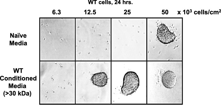
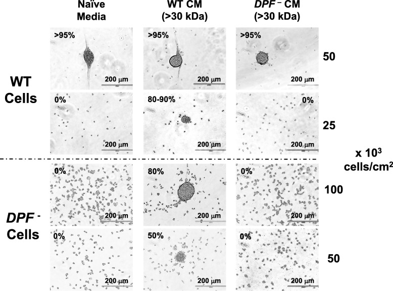
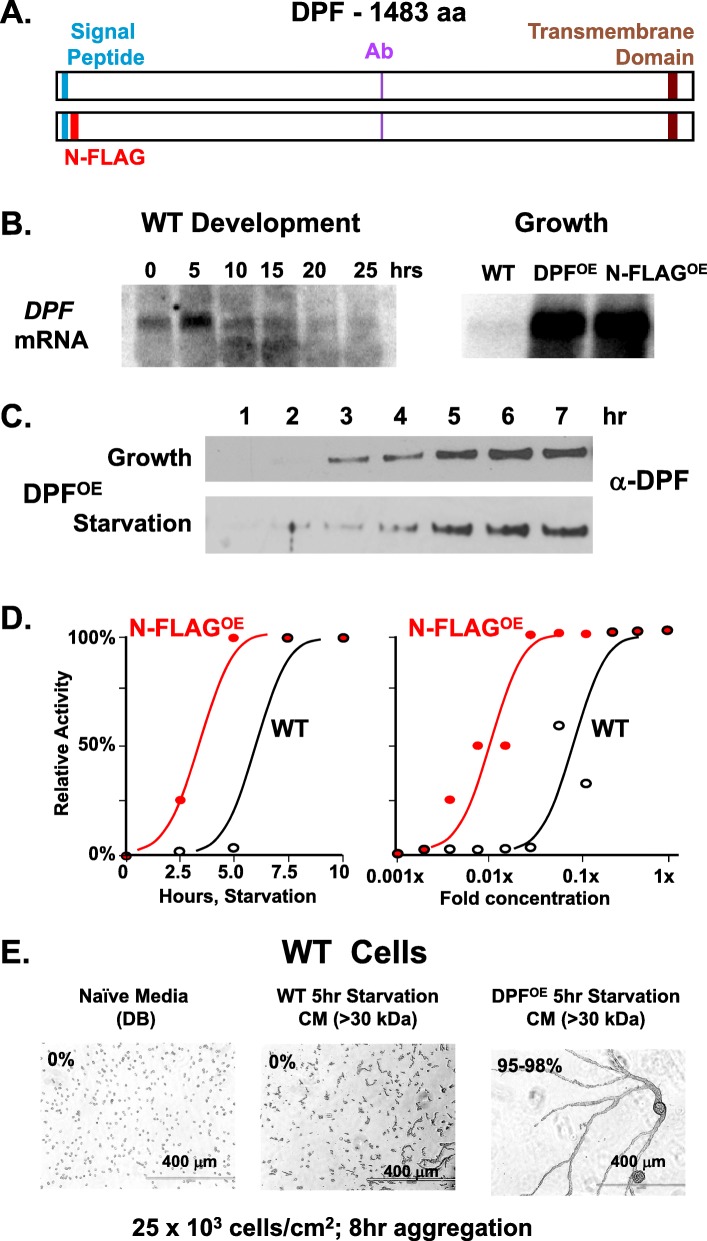
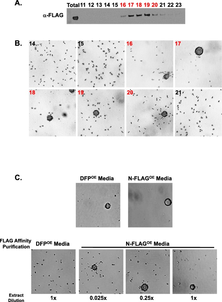
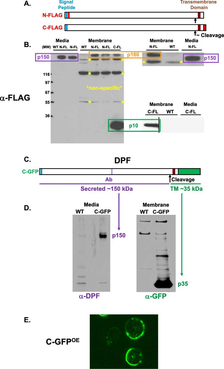
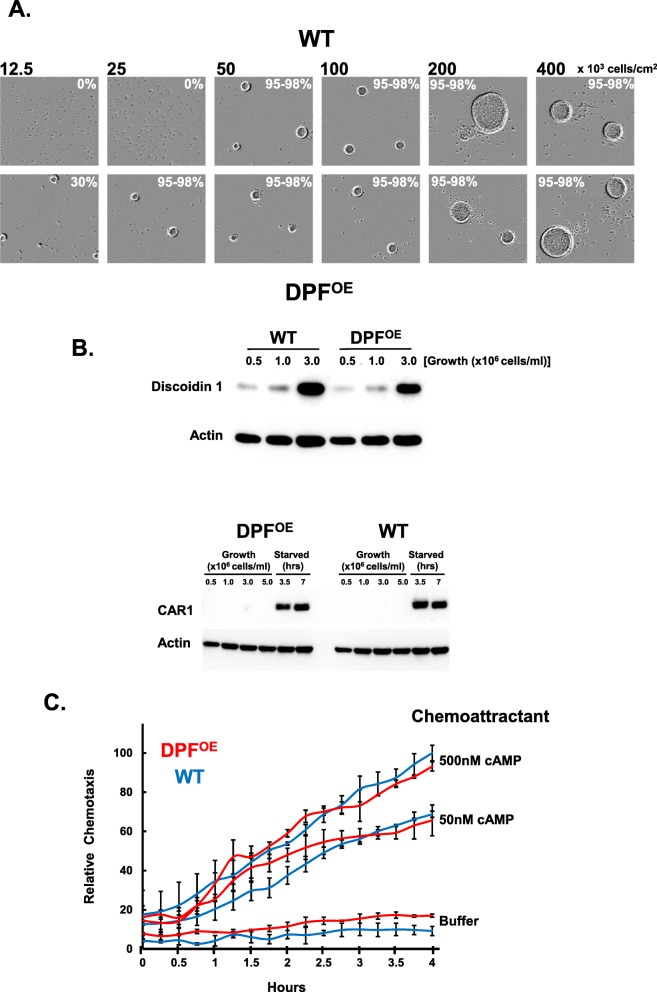
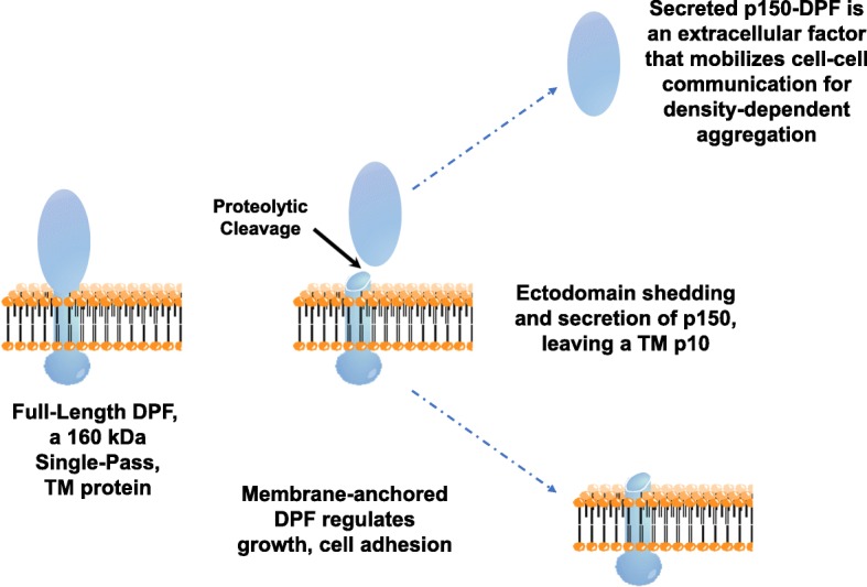
Results: We show that multi-cell developmental aggregation in Dictyostelium is lost upon minimal (2-fold) reduction in local cell density. Remarkably, developmental aggregation response at non-permissive cell densities is rescued by addition of conditioned media from high-density, developmentally competent cells. Using rescued aggregation of low-density cells as an assay, we purified a single, 150-kDa extra-cellular protein with density aggregation activity. MS/MS peptide sequence analysis identified the gene sequence, and cells that overexpress the full-length protein accumulate higher levels of a development promoting factor (DPF) activity than parental cells, allowing cells to aggregate at lower cell densities; cells deficient for this DPF gene lack density-dependent developmental aggregation activity and require higher cell density for cell aggregation compared to WT. Density aggregation activity co-purifies with tagged versions of DPF and tag-affinity-purified DPF possesses density aggregation activity. In mixed development with WT, cells that overexpress DPF preferentially localize at centers for multi-cell aggregation and define cell-fate choice during cytodifferentiation. Finally, we show that DPF is synthesized as a larger precursor, single-pass transmembrane protein, with the p150 fragment released by proteolytic cleavage and ectodomain shedding. The TM/cytoplasmic domain of DPF possesses cell-autonomous activity for cell-substratum adhesion and for cellular growth.

Conclusions: We have purified a novel secreted protein, DPF, that acts as a density-sensing factor for development and functions to define local collective thresholds for Dictyostelium development and to facilitate cell-cell communication and multi-cell formation. Regions of high DPF expression are enriched at centers for cell-cell signal-response, multi-cell formation, and cell-fate determination. Additionally, DPF has separate cell-autonomous functions for regulation of cellular adhesion and growth.

Keywords: Chemotaxis; Ecto-domain shedding; MS/MS peptide sequencing; Protein purification; Signaling.


Conflict of interest statement

The authors declare that they have no competing interests.

Figures

Fig. 1
Fig. 1
Conditioned media promotes aggregation at low cell density. Log phase growing WT cells were adhered in a 12-well plate under DB starvation buffer at indicated cell densities for 24 h, using either fresh, naïve DB media, or cell-free, > 30-kDa conditioned media from WT cells starved in DB for 18 h (see also Additional file 1: Figure S1A,B)
Fig. 2
Fig. 2
Purification of the density aggregation activity. a The cell-free, > 30-kDa conditioned media from WT cells were fractionated on mono Q, phenyl sepharose (PS), wheat germ agglutinin (WG), and Superose 12 columns. Fractions were assayed for density-dependent aggregation activity on WT cells at low cell density (< 20 × 103 cells/cm2). Selected bound or flow through (FT) fractions separated by SDS gel electrophoresis are shown. Gels were stained with silver and protein bands indicated from Superose 12 fractions (e.g., 11 and 12) were used for peptide sequencing. Proteins matching each band are indicated (see Additional file 3: Figure S3). b The procedure followed Fig. 2a, but using conditioned DB from PDE1-null cells. Superose 12 fractions 22, 23, and 24 were used for MS/MS peptide sequencing. c Comparison of Superose 12 fractionations of conditioned DB from WT or PDE1-nulls cells, with protein profiles, relative MW positions, and activity position shift indicated. Relative fractionation differences for the proteins in Fig. 2a are also shown
Fig. 3
Fig. 3
Cells deficient in p150 lack aggregation promoting activity. Log phase growing WT or DPF-null cells were adhered in a 12-well plate under DB starvation buffer at indicated cell densities for 24 h, using either fresh, naïve DB media or cell-free, > 30-kDa conditioned media from WT or DPF-null cells starved in DB for 18 h. Relative density-dependent activity for each is indicated as % aggregation. Scale bar = 200 μm
Fig. 4
Fig. 4
p150 protein structure, as termed DPF, and expression patterns. a Predicted structure of protein p150 (DPF). Full-length DPF has an N-terminal signal peptide and C-terminal transmembrane domain. An antibody was to a specific peptide (Additional file 3: Figure S3C). The relative positions these features are positioned along the 1483 amino acid backbone. The N-FLAG DPF expression construct was created with a FLAG peptide sequence inserted in-frame, 3′ to the signal peptide. b Left panel—developmental expression of DPF mRNA at indicated times for WT cells, using RNA-blot hybridization. Right panel—relative DPF mRNA levels in growing WT cells, WT cells expressing full-length DPF (DPFOE), or WT cells expressing full-length FLAG-tagged DPF (N-FLAGOE), using RNA-blot hybridization. c Log-phase growing WT cells overexpressing DPF (DPFOE) were transferred into fresh growth media or fresh DB starvation buffer at similar cell densities and supernatant fractions taken and immunoblotted to α-DPF (see Fig. 4a). d Left panel—growing WT or WT cells expressing full-length FLAG-tagged DPF (N-FLAGOE) were transferred into fresh DB. Cell-free media were taken at times indicated and tested for relative density-dependent aggregation activity, using WT cells at 20 × 103 cells/cm2. Right panel—media collected at 7.5 h from both WT or N-FLAGOE cells were diluted into fresh DB, as indicated, and tested for relative density-dependent aggregation activity, using WT cells at 20 × 103 cells/cm2. Starting medium (1×) is 10-fold diluted, from a centricon concentrate of conditioned supernatant. e Log-phase growing WT cells were plated under DB buffer at 25 × 103 cells/cm2 for 8 h, using either fresh, naïve DB media, or cell-free, > 30-kDa conditioned media from WT or DPFOE cells starved in DB for 5 h (see Fig. 4d,e). Relative density-dependent activity is indicated as % aggregation. Scale = 400 μm
Fig. 5
Fig. 5
FLAG-p150 co-purifies with density aggregation activity. a/b Conditioned media from N-FLAGOE cells was fractionated on mono Q and eluate fractions assayed by immunoblot for FLAG protein (a) and for density-dependent aggregation activity (b) using WT cells at 20 × 103 cells/cm2. c Upper panels—Mono Q fractionated media from DPFOE or N-FLAGOE cells were assayed for density-dependent aggregation activity using WT cells at 20 × 103 cells/cm2. Lower panels—media were affinity purified with α-FLAG and re-assayed at varying dilutions for density-dependent aggregation activity using WT cells at 20 × 103 cells/cm2
Fig. 6
Fig. 6
p150 is released from the plasma membrane by ectodomain shedding. a Two DPF constructs were engineered. One has an N-terminal FLAG (see Fig. 4a) and the other a C-terminal FLAG. b Cells expressing N-FLAG and C-FLAG were shaken in DB for 18 h and media and membrane fractions prepared and immunoblotted to α-FLAG. The most abundant N- and C-terminal tags are localized to separate sized fragments, suggesting processed cleavage for ectodomain shedding. A full-length DPF form is in the membrane as p160; it is processed to release p150 and membrane-anchored p10 (see Fig. 6a). c A C-terminal GFP DPF protein expression construct was also engineered. d Media and membrane fractions from WT cells and WT cells expressing C-GFP (C-GFPOE). Media proteins were immunoblotted to α-DPF (see Fig. 4a,c and Additional file 3: Figure S3C), and membrane proteins were immunoblotted to α-GFP. α-DPF detects secreted p150 and α-GFP detects membrane-anchored p10 fused to GFP. e Fluorescence localization of GFP in C-GFP expressing cells. Strong GFP fluorescence, as a read-out of the DPF TM domain, is seen at the cell periphery
Fig. 7
Fig. 7
Gain-of-function studies of WT cells expressing high levels of DPF. a Log phase growing WT or DPFOE cells were plated under-buffer at indicated cell densities for 24 h using fresh, naïve DB media. Relative aggregation efficiencies are indicated. b WT and DPFOE, DPF-OE cells were grown to various cell densities in growth media and then identically starved as indicated. Cell lysates were prepared from cells at indicated times and immunoblotted to α-Discoidin 1, α-CAR1, and α-actin. c Time-course quantification of WT or DPFOE cell migration to various doses of cAMP. Relative chemotaxis is normalized to WT cells at 500 nM cAMP at 4 h. Standard deviations are shown based upon three replicates
Fig. 8
Fig. 8
Cells that overexpress DPF regulate prespore/spore patterning. a WT or C-GFPOE cells were identically plated on DB agar at a density of 400 × 103 cells/cm2 for development and followed over time. Shown are similar time frame images including both DIC and GFP fluorescence. b A 9:1 mixed population of WT or C-GFPOE cells were plated for development and followed over time. Shown are two time frame images including both DIC and GFP fluorescence (see Additional file 8: Movie S1). c A 99:1 mixed population of WT or C-GFPOE cells were plated for development and developed to the slug stage (left) or to terminal differentiation (right). Shown are confocal images including both DIC and GFP fluorescence, with prespore/prestalk and spore/stalk regions indicated
Fig. 9
Fig. 9
Cell-autonomous and non-autonomous functions of DPF in development. a Log phase growing DPF- cells were adhered in a 12-well plate under fresh, naïve DB starvation buffer at indicated cell densities for 24 h. b Log phase growing DPF- or DPF-OE Dictyostelium were plated under fresh, naïve DB starvation buffer at indicated cell densities for 24 h. c WT, DPF-, DPFOE, and DPF-OE cells were adhered in fresh, naïve DB media at a density of 100 × 103 or 400 × 103 cells/cm2 and developed for 5 h. Cell lysates were prepared and immunoblotted to α-CAR1 and α-actin. d WT and DPF- cells were adhered at a density of 100 × 103 cells/cm2 and developed for 5 h using fresh, naïve DB media, or > 30-kDa conditioned media from WT, DPFOE, or DPF- cells following starvation in DB for 5 h. Cell lysates were prepared and immunoblotted to α-CAR1 and α-actin
Fig. 10
Fig. 10
Cell-autonomous functions of DPF in adhesion and growth. a WT and DPF- cells were adhered at a density of 400 × 103 cells/cm2 to a six-well plate, washed and replenished with fresh, naïve DB media or 5-h conditioned media from DPFOE cells. The dishes were then shaken at indicated time points at 90 rpm, and the percentage of de-attached cells quantified. Values indicate Mean ± SD from triplicate sets and three independent experiments. b Cell growth rates of WT and DPF- cells in the presence of fresh growth media that was supplemented with DPF-containing conditioned growth media from WT cells. Growth rate was monitored at indicated time points. The values represent mean ± SD from three independent experiments
Fig. 11
Fig. 11
A model for ectodomain shedding and secretion of DPF. DPF is synthesized as an ~ 160-kDa protein that is inserted into the plasma membrane, following signal peptide cleavage. The transmembrane TM domain near the C-terminus anchors the single-pass DPF in the membrane. The long N-terminal ~ 150-kDa extracellular domain is glycosylated. Extracellular proteolytic cleavage, N-terminal to the TM domain, releases a p150 fragment into the media; The residual p10 TM/cytoplasmic fragment is retained in the plasma membrane. The secreted p150 possesses density-dependent aggregation activity and at high levels promotes aggregation at sub-optimal cellular densities and defines centers for aggregation. Membrane-anchored DPF has cell-autonomous activity for growth and adhesion

Similar articles

Cited by

References

    1. Abisado RG, Benomar S, Klaus JR, Dandekar AA, Chandler JR. Bacterial quorum sensing and microbial community interactions. mBio. 2018;9(3):e02331–e02317. doi: 10.1128/mBio.02331-17. - DOI - PMC - PubMed
    1. Polonsky M, Rimer J, Kern-Perets A, Zaretsky I, Miller S, Bornstein C, David E, Kopelman NM, Stelzer G, Porat Z, et al. Induction of CD4 T cell memory by local cellular collectivity. Science. 2018;360(6394):eaaj1853. doi: 10.1126/science.aaj1853. - DOI - PubMed
    1. Watt KI, Harvey KF, Gregorevic P. Regulation of tissue growth by the mammalian hippo signaling pathway. Front Physiol. 2017;8:942. doi: 10.3389/fphys.2017.00942. - DOI - PMC - PubMed
    1. Sagner Andreas, Briscoe James. Morphogen interpretation: concentration, time, competence, and signaling dynamics. Wiley Interdisciplinary Reviews: Developmental Biology. 2017;6(4):e271. doi: 10.1002/wdev.271. - DOI - PMC - PubMed
    1. Eder D, Aegerter C, Basler K. Forces controlling organ growth and size. Mech Dev. 2017;144(Pt A):53–61. doi: 10.1016/j.mod.2016.11.005. - DOI - PubMed

Publication types

MeSH terms

Substances

LinkOut - more resources