. 2019 Dec 2;9(1):18110.
doi: 10.1038/s41598-019-54711-z.

Plug-and-Play In Vitro Metastasis System toward Recapitulating the Metastatic Cascade

Affiliations

Plug-and-Play In Vitro Metastasis System toward Recapitulating the Metastatic Cascade

Bing-Syuan Ni et al. Sci Rep. .

Abstract

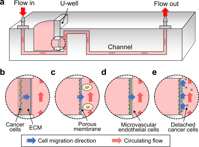
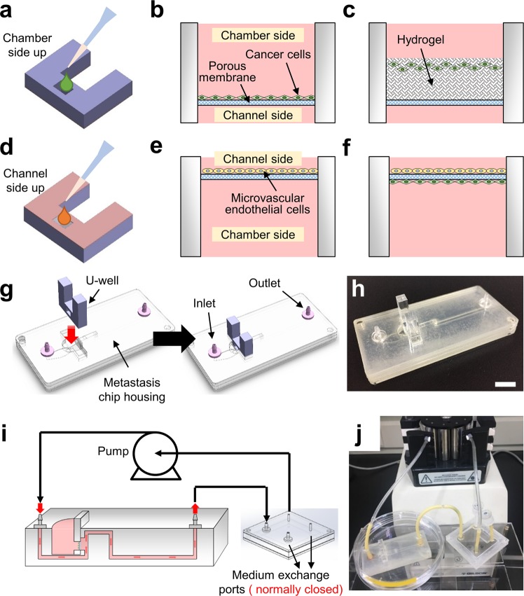
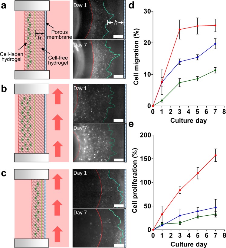
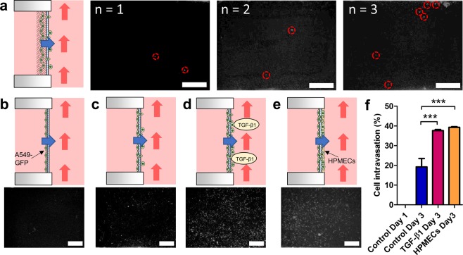
Microfluidic-based tumor models that mimic tumor culture environment have been developed to understand the cancer metastasis mechanism and discover effective antimetastatic drugs. These models successfully recapitulated key steps of metastatic cascades, yet still limited to few metastatic steps, operation difficulty, and small molecule absorption. In this study, we developed a metastasis system made of biocompatible and drug resistance plastics to recapitulate each metastasis stage in three-dimensional (3D) mono- and co-cultures formats, enabling the investigation of the metastatic responses of cancer cells (A549-GFP). The plug-and-play feature enhances the efficiency of the experimental setup and avoids initial culture failures. The results demonstrate that cancer cells tended to proliferate and migrate with circulating flow and intravasated across the porous membrane after a period of 3 d when they were treated with transforming growth factor-beta 1 (TGF-β1) or co-cultured with human pulmonary microvascular endothelial cells (HPMECs). The cells were also observed to detach and migrate into the circulating flow after a period of 20 d, indicating that they transformed into circulating tumor cells for the next metastasis stage. We envision this metastasis system can provide novel insights that would aid in fully understanding the entire mechanism of tumor invasion.


Conflict of interest statement

The authors declare no competing interests.

Figures

Figure 1
Figure 1
(a) Schematic diagram illustrates a cross-section view of the metastasis chip. The design allows for the evaluation of the metastasis capability of cancer cells at different stages using a plug-and-play design. The U-well can be removed and placed under a microscope to monitor the cell behaviors inside the region of the U-well circled with a dashed line: (b) Cancer cell migration through the ECM. (c) Cancer cell intravasation using growth factor (GF). (d) Cancer cell intravasation with a co-culture of microvascular endothelial cells. (e) Cancer cell detachment into the circulating flow.
Figure 2
Figure 2
U-well and plug-and-play culture design allowing for culturing various types of cancer cells and performing diverse culture techniques in a simple fashion. (a) The cancer cells can be directly seeded on the chamber side of the U-well using a pipette. (b) Once the cancer cells are seeded, these cells can attach to the porous membrane to form a mono-cell layer after incubation. (c) The cell-laden hydrogel can be pre-mixed with and loaded into the U-well to generate a 3D culture environment. (d) The microvascular endothelial cells can also be seeded on the channel side of the U-well to investigate the influence of shear stress. (e) The attached microvascular endothelial cells on the channel side of the U-well. (f) The cancer cells can be seeded before or after the seeding of microvascular endothelial cells to form a co-culture environment. (g) The U-well can be plugged into the metastasis chip housing directly to complete the metastasis chip. The inlet and outlet of the chip housing are connected to the perfusion system. (h) An image of the metastasis chip. Scale bar = 1 cm. (i) Schematic setup of the metastasis system with a collection device and a peristaltic pump allowing for the collection of the circulating tumor cells under dynamic flow conditions. (j) An image of metastasis system setup.
Figure 3
Figure 3
A549-GFP cell migration in the chip. (a) Cells migrate under static flow conditions through the cell-free hydrogel with a controlled thickness h ~ 765 µm. The red dashed line indicates the boundary between the cell-laden hydrogel and culture medium. The green line represents the leading front of the cells. Initially, it indicates the boundary between the cell-free and cell-laden hydrogels on day 0. Fluorescent images demonstrate the cells migration and proliferation after 1 and 7 days of culture. (b) Cells migrating through the thick cell-free hydrogel (h ~ 765 µm) under flow conditions. The red arrow indicates the flow direction. (c) Cells migrating through the thin cell-free hydrogel (h ~ 160 µm) under flow conditions. Scale bar = 500 μm. (d) Cell migration percentage under various conditions. Cells migrated through a thick hydrogel with static culture conditions (green), through a thick hydrogel with dynamic culture conditions (blue), and through a thin hydrogel with dynamic culture conditions (red). (e) Cell proliferation percentage under different culture conditions. Cell proliferated in a thick hydrogel with static culture conditions (green), in a thick hydrogel with dynamic culture conditions (blue), and in a thin hydrogel with dynamic culture conditions (red). N = 3.
Figure 4
Figure 4
Intravasation of A549-GFP cells across the membrane in the chip. (a) Cell migration and intravasation from the collagen to the channel side of the U-well after 7 days of culture. Similar results were observed in three repeated experiments under a fluorescence microscope. Intravasated cells are enclosed in red circles. (b) A549-GFP cells were cultured in a monolayer condition for 1 d. The fluorescent image demonstrates no cells intravasated across the membrane. (c) A549-GFP cells were cultured for 3 d and began to intravasate. (d) A549-GFP cells were cultured for 3 d with TGF-β1 treatment, triggering more cells to intravasated. (e) A549-GFP cells were co-cultured with HPMECs for 3 d, enhancing the cell intravasation. (f) Cell intravasated percentages for the A549-GFP in the metastasis chip. ***P < 0.001. All cells on the chamber side of the U-well were removed to avoid the interference. Scale bar = 500 μm.
Figure 5
Figure 5
Dissociation of A549-GFP cells from the primary site. (a) The schematic demonstrates the intravasated cells detaching from the porous membrane and migrating with the circulating medium flow. (b) The cell collector connected after the metastasis chip can ensure the collection of dissociated cells. The sedimentation locations of detached cells in the cell collector were imaged after 20 days of culture. Cells are circled in green. Scale bar = 500 μm.

References

    1. Chaffer CL, Weinberg RA. A perspective on cancer cell metastasis. Science. 2011;331:1559–1564. doi: 10.1126/science.1203543. - DOI - PubMed
    1. Lambert AW, Pattabiraman DR, Weinberg RA. Emerging Biological Principles of Metastasis. Cell. 2017;168:670–691. doi: 10.1016/j.cell.2016.11.037. - DOI - PMC - PubMed
    1. Gandalovicova A, et al. Migrastatics-Anti-metastatic and Anti-invasion Drugs: Promises and Challenges. Trends Cancer. 2017;3:391–406. doi: 10.1016/j.trecan.2017.04.008. - DOI - PMC - PubMed
    1. Fernandes M, Rosel D, Brabek J. Translation in solid cancer: are size-based response criteria an anachronism? Clin Transl Oncol. 2015;17:1–10. doi: 10.1007/s12094-014-1207-5. - DOI - PubMed
    1. Hosseini H, et al. Early dissemination seeds metastasis in breast cancer. Nature. 2016;540:552–558. doi: 10.1038/nature20785. - DOI - PMC - PubMed

Publication types