Case Reports
. 2020 Apr;28(4):445-452.
doi: 10.1038/s41431-019-0546-7. Epub 2019 Dec 2.

Biallelic variants in EFEMP1 in a man with a pronounced connective tissue phenotype

Affiliations
Case Reports

Biallelic variants in EFEMP1 in a man with a pronounced connective tissue phenotype

Sean G W Driver et al. Eur J Hum Genet. 2020 Apr.

Abstract

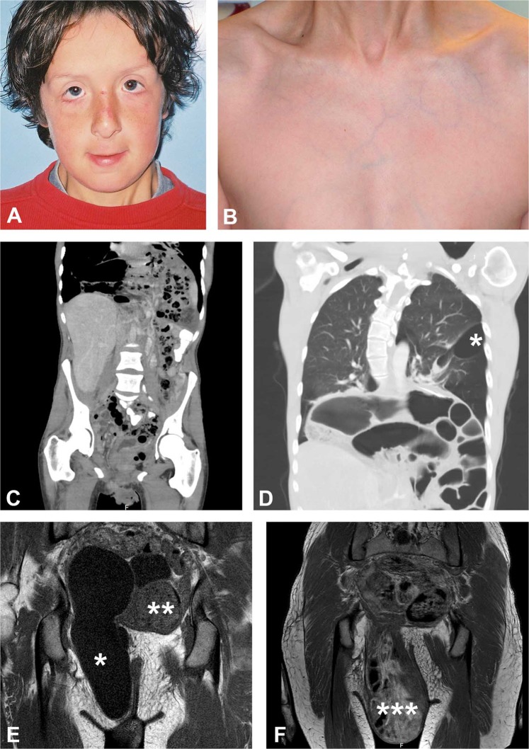
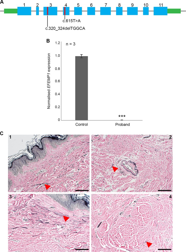
Connective tissue disorders are a spectrum of diseases that affect the integrity of tissues including skin, vasculature, and joints. They are often caused by variants that disrupt genes encoding components of extracellular matrix (ECM). The fibulin glycoproteins are ECM proteins important for integrity of tissues including dermis, retina, fascia, and vasculature. The fibulin family consists of seven members (fibulins-1 to -7) and is defined by a fibulin-type domain at the C-terminus. The family is associated with human diseases, for instance a variant in FBLN1, encoding fibulin-1, is associated with synpolydactyly, while one in EFEMP1, encoding fibulin-3, causes Doyne honeycomb degeneration of the retina. Loss-of-function of fibulins-4 and -5 causes cutis laxa, while variants in fibulins-5 and -6 are associated with age-related macular degeneration. Of note, EFEMP1 is not currently associated with any connective tissue disorder. Here we show biallelic loss-of-function variants in EFEMP1 in an individual with multiple and recurrent abdominal and thoracic herniae, myopia, hypermobile joints, scoliosis, and thin translucent skin. Fibroblasts from this individual express significantly lower EFEMP1 transcript than age-matched control cells. A skin biopsy, visualised using light microscopy, showed normal structure and abundance of elastic fibres. The phenotype of this individual is remarkably similar to the Efemp1 knockout mouse model that displays multiple herniae with premature aging and scoliosis. We conclude that loss of EFEMP1 function in this individual is the cause of a connective tissue disorder with a novel combination of phenotypic features, and can perhaps explain similar, previously reported cases in the literature.


Conflict of interest statement

The authors declare that they have no conflict of interest.

Figures

Fig. 1
Fig. 1
Clinical images of the proband. a Distinctive facial features include a long, narrow face, ectropia of the lower eyelids, and downslanting palpebral fissures; b The skin is thin and translucent; c Large Bochdalek hernia on both sides of the mediastinum with loops of small bowel herniated into the thorax, along with spleen and mesenteric vessels. An obturator hernia is also evident on the left with bladder content; d Diaphragmatic hernia with small bowel loops and stomach in the thorax with multiple pleural based bullae (*) throughout both lung fields; e Coronal MRI of the pelvis with a bladder diverticulum (*) and left obturator hernia (**) containing bladder evident; f Coronal MRI of the pelvis with a pelvic floor hernia with small bowel loop plus bladder content present (***) in addition to a small left obturator hernia
Fig. 2
Fig. 2
a EFEMP1 gene structure (NM_001039348.3), variants discovered in this study are highlighted, numbers represent exons; b qPCR data showed a significant reduction in EFEMP1 expression from affected individual’s fibroblasts compared with those from an age- and sex-matched control. EFEMP1 expression was normalised to GAPDH (***p < 0.001); c Panels 1–4; healthy skin biopsy sections taken from the affected individual, treated with Verhoeff–van Gieson (VVG) to stain the elastin fibres. Elastin is stained black. At ×20 magnification, the elastic fibres appear normal and correctly distributed (arrows). Scale bars represent 100 µm (Color figure online)

References

    1. Milewicz DM, Pyeritz RE, Crawford ES, Byers PH. Marfan syndrome: defective synthesis, secretion, and extracellular matrix formation of fibrillin by cultured dermal fibroblasts. J Clin Investig. 1992;89:79–86. doi: 10.1172/JCI115589. - DOI - PMC - PubMed
    1. Dietz HC, Cutting CR, Pyeritz RE, Maslen CL, Sakai LY, Corson GM, et al. Marfan syndrome caused by a recurrent de novo missense mutation in the fibrillin gene. Nature. 1991;352:337–9. doi: 10.1038/352337a0. - DOI - PubMed
    1. Beighton P, Paepe AD, Steinmann B, Tsipouras P, Wenstrup RJ. Ehlers-Danlos syndromes: revised nosology. Am J Med Genet. 1998;77:31–37. doi: 10.1002/(SICI)1096-8628(19980428)77:1<31::AID-AJMG8>3.0.CO;2-O. - DOI - PubMed
    1. Malfait F, de Paepe A. Molecular genetics in classic Ehlers–Danlos syndrome. Am J Med Genet Part C Semin Med Genet. 2005;139C:17–23. doi: 10.1002/ajmg.c.30070. - DOI - PubMed
    1. Pepin M, Schwarze U, Superti-Furga A, Byers PH. Clinical and genetic features of Ehlers–Danlos syndrome type IV, the vascular type. N Engl J Med. 2000;342:673–80. doi: 10.1056/NEJM200003093421001. - DOI - PubMed

Publication types

Substances