Comparative Study
. 2019 Dec;26(12):1151-1157.
doi: 10.1038/s41594-019-0334-7. Epub 2019 Dec 2.

Structures of MERS-CoV spike glycoprotein in complex with sialoside attachment receptors

Affiliations
Comparative Study

Structures of MERS-CoV spike glycoprotein in complex with sialoside attachment receptors

Young-Jun Park et al. Nat Struct Mol Biol. 2019 Dec.

Abstract

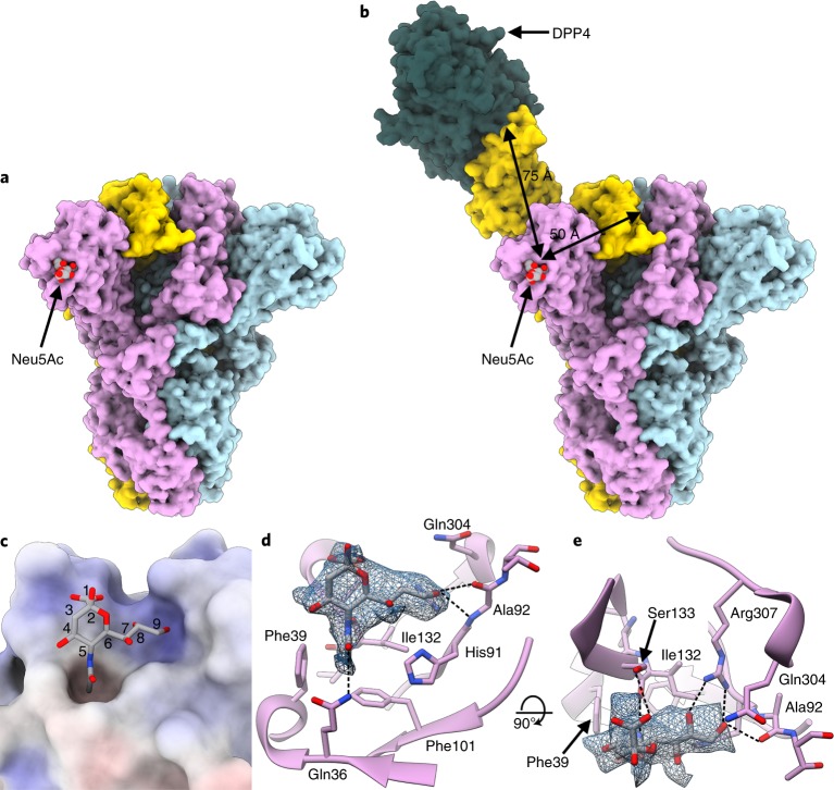
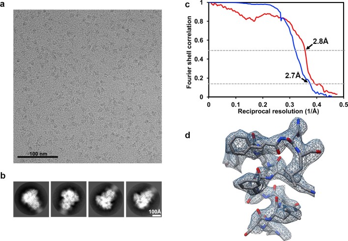
The Middle East respiratory syndrome coronavirus (MERS-CoV) causes severe and often lethal respiratory illness in humans, and no vaccines or specific treatments are available. Infections are initiated via binding of the MERS-CoV spike (S) glycoprotein to sialosides and dipeptidyl-peptidase 4 (the attachment and entry receptors, respectively). To understand MERS-CoV engagement of sialylated receptors, we determined the cryo-EM structures of S in complex with 5-N-acetyl neuraminic acid, 5-N-glycolyl neuraminic acid, sialyl-LewisX, α2,3-sialyl-N-acetyl-lactosamine and α2,6-sialyl-N-acetyl-lactosamine at 2.7-3.0 Å resolution. We show that recognition occurs via a conserved groove that is essential for MERS-CoV S-mediated attachment to sialosides and entry into human airway epithelial cells. Our data illuminate MERS-CoV S sialoside specificity and suggest that selectivity for α2,3-linked over α2,6-linked receptors results from enhanced interactions with the former class of oligosaccharides. This study provides a structural framework explaining MERS-CoV attachment to sialoside receptors and identifies a site of potential vulnerability to inhibitors of viral entry.


Conflict of interest statement

The authors declare no competing interests.

Figures

Fig. 1
Fig. 1. Cryo-EM identification of a sialoside-binding site in the MERS-CoV S glycoprotein.
a, Molecular surface representation of the MERS-CoV S ectodomain trimer, with each protomer in a different color. The Neu5Ac ligands are rendered as spheres. b, Spatial relationships between the attachment (sialoside) and entry (DDP4) receptor-binding sites are illustrated using a composite model obtained from the cryo-EM structure shown in a, the cryo-EM structure of MERS-CoV S bound to the LCA60 neutralizing antibody Fab fragment (PDB 6NB3) and a crystal structure of the MERS-CoV S domain B bound to DPP4 (PDB 4KR0). The sialoside-binding site is located ~50 and ~75 Å away from the DPP4-binding site of a neighboring B domain in the closed or open state, respectively. c, Surface representation of the ligand-binding site colored by electrostatic potential from −12 (red) to +10 (blue) kBT/ec. Neu5Ac is shown in stick representation. d,e, Two orthogonal views of the sialoside-binding site rendered as a ribbon diagram with the side chains of key surrounding residues shown as sticks. Neu5Ac is rendered as sticks and the corresponding region of cryo-EM density is shown as a blue mesh contoured at 5.5σ. Dashed lines show selected electrostatic interactions formed between MERS-CoV S amino acid residues and the ligand. In all panels, nitrogen and oxygen atoms are colored blue and red, respectively, while carbon atoms are colored gray (Neu5Ac) or pink (MERS-CoV S). Glycans in MERS-CoV S have been omitted for clarity.
Fig. 2
Fig. 2. MERS-CoV S and HCoV-OC43 S glycoproteins interact with sialosides, using distinct binding grooves.
a,b, Molecular surface representation of the MERS-CoV S (a) and HCoV-OC43 S (b) A domains bound to Neu5Ac and 5-N-acetyl,9-O-acetyl neuraminic acid α-methyl glycoside (9-O-Ac-Neu5Ac, PDB 6NZK), respectively, and oriented identically. The sialosides are rendered as sticks with carbon, nitrogen and oxygen atoms colored gray, blue and red, respectively. N-linked glycans observed in the cryo-EM reconstructions are rendered as dark blue spheres. c, Ribbon diagrams of superimposed MERS-CoV S (pink) and HCoV-OC43 S (gold) A domains with bound sialosides. The sialosides are rendered as sticks with carbon, nitrogen and oxygen atoms colored pink (MERS-CoV S) or gold (HCoV-OC43 S), blue and red, respectively.
Fig. 3
Fig. 3. The ligand-binding site is required for MERS-CoV S-mediated attachment to sialosides and entry into human airway epithelial cells.
a, Hemagglutination assay using MERS-CoV S wild type (WT) or mutants. Wells positive for hemagglutination (HA) are encircled. F39A, H91A, I132A, S133A or R307A substitutions completely abrogated domain A-Fc-mediated hemagglutination of human erythrocytes. Mock-treated erythrocytes, absence of the protein A-fused lumazine synthase (pA-LS) or replacement of domain A with domain B (Dom. B) were used as negative controls. The assays were performed four times and a representative experiment is shown. b, MERS-CoV S F39A, H91A, S133A or R307A substitutions inhibited entry of pseudotyped murine leukemia viral particles into human airway Calu-3 cells. Data are normalized relative to wild type and shown as mean and s.d. of n = 3 pseudovirus experiments (technical replicates).
Fig. 4
Fig. 4. Structural basis of MERS-CoV S selectivity for α2,3-linked sialoside receptors.
a–f, Enlarged views of the MERS-CoV S structures in complex with sLeX (a,b), 2,3-SLN (c,d) and 2,6-SLN (e,f). The cryo-EM densities corresponding to the ligands are shown as a blue mesh contoured at 3.7σ (a), 3.2σ (b), 5.8σ (c,d) and 4.5σ (e,f). g,h, The ligand-binding site with the sLeX and 2,6-SLN-bound MERS-CoV S structures superimposed, showing the distinct orientation of the two ligands relative to domain A. The asterisks indicate the proximity of the sLeX GlcNAc to the binding site, whereas the 2,6-SLN GlcNAc points away towards the solvent. In ad, the A domain is rendered as a ribbon diagram with selected side chains of residues surrounding the sialoside shown as sticks. The ligands are rendered as sticks with atoms colored by elements (carbon, cyan (sLeX), green (2,3-SLN) or orange (2,6-SLN); nitrogen, blue; oxygen, red). Neu5Ac, 5-N-acetyl neuraminic acid; GlcNAc, N-acetyl glucosamine; Fuc, fucose; Gal, galactose.
Extended Data Fig. 1
Extended Data Fig. 1. Chemical structures of the sialosides used for structural studies.
a, Neu5Ac. b, Neu5Gc. c, sLeX. d, 2,3-SLN. e, 2,6-SLN.
Extended Data Fig. 2
Extended Data Fig. 2. CryoEM analysis of the MERS-CoV S glycoprotein in complex with Neu5Ac at 2.7 Å resolution.
a,b, Representative electron micrograph (a) and class averages (b) for the Neu5Ac-bound MERS-CoV S structure. c, Gold-standard (blue) and map/model (red) Fourier shell correlation curves. The 0.143 and 0.5 cutoffs are indicated by horizontal dashed lines. d, Local resolution map calculated using cryoSPARC. The Neu5Ac ligand is estimated to be resolved at 3Å resolution. eg, Representative cryoEM densities shown as blue mesh with the corresponding atomic model rendered as sticks colored grey, blue and red for carbon, nitrogen and oxygen atoms, respectively. Dashed bonds indicate hydrogen bonds with ordered water molecules.
Extended Data Fig. 3
Extended Data Fig. 3. Structural basis for the MERS-CoV S selectivity for neuraminic acids that are not 9-O-acetylated or 5-N-glycolylated.
a, Superimposition of the 5-N-acetyl,9-O-acetyl neuraminic acid α-methyl glycoside (9-O-Ac-Neu5Ac, green) from the HCoV-OC43 S holo structure (PDB 6NZK) to the bound Neu5Ac from the MERS-CoV S/Neu5Ac complex structure suggests the MERS-CoV S sialoside-binding site could not sterically accommodate the 9-O-acetyl group in this conformation. The distances between the 9-O-acetyl group and His91 or Ala92 are indicated. b, The MERS-CoV S sialoside-binding site rendered as a ribbon diagram with the side chains of key surrounding residues shown. Neu5Ac is rendered with the corresponding region of cryoEM density from the MERS-CoV S/Neu5Ac complex structure (low-pass filtered to 3Å and scaled to the MERS-CoV S/Neu5Gc map) shown as a blue mesh contoured at 6σ. cf, The cryoEM structure of MERS-CoV S in complex with Neu5Gc shows weaker density for the sialoside (blue mesh contoured at 6σ), indicating poor steric and/or chemical accommodation of the 5-N-glycolyl hydroxyl group in the hydrophobic pocket defined by Phe39, Phe101, Ile131 and Ile132. In panels (c-e) Neu5Gc was modeled based on the porcine rotavirus CRW-8 VP8* holo structure (PDB 3TAY) which corresponds to the conformation observed in 1 out of 8 structures available in the protein data bank with a Neu5Gc analogue. In panel f, Neu5Gc was modeled based on the rhesus rotavirus VP8* holo structure (PDB 3TB0) which is the only structure in the protein data bank featuring Neu5Gc with the 5-N-glycolyl group in an alternate orientation. The distance between the 5-N-glycolyl hydroxyl group and Gln36 is indicated.
Extended Data Fig. 4
Extended Data Fig. 4. CryoEM analysis of the MERS-CoV S glycoprotein in complex with Neu5Gc at 3.0 Å resolution.
a,b, Representative electron micrograph (a) and class averages (b) for the Neu5Gc-bound MERS-CoV S structure. c, Gold-standard Fourier shell correlation curve. The 0.143 cut-off is indicated by horizontal dashed lines. d, Representative density shown as blue mesh with the corresponding atomic model rendered as sticks colored grey, blue and red for carbon, nitrogen and oxygen atoms, respectively.
Extended Data Fig. 5
Extended Data Fig. 5. SDS-PAGE and Western blot analyses.
a, SDS-PAGE analysis of purified wild type or mutants MERS-CoV S domain A fused to human immunoglobulin Fc. Two micrograms of each protein was loaded. b, Western-blot analysis of murine leukemia viral particles pseudotyped with wild type or mutants MERS-CoV S using an anti-MERS-CoV S1 polyclonal antibody. Uncropped blot image is available as source data. Source data
Extended Data Fig. 6
Extended Data Fig. 6. CryoEM analysis of the MERS-CoV S glycoprotein in complex with sLeX at 2.7 Å resolution.
a,b, Representative electron micrograph (a) and class averages (b) for the sLeX-bound MERS-CoV S structure. c, Gold-standard (blue) and map/model (red) Fourier shell correlation curves. The 0.143 and 0.5 cutoffs are indicated by horizontal dashed lines. d, Representative density shown as blue mesh with the corresponding atomic model rendered as sticks colored grey, blue and red for carbon, nitrogen and oxygen atoms, respectively.
Extended Data Fig. 7
Extended Data Fig. 7. CryoEM analysis of the MERS-CoV S glycoprotein in complex with 2,3-SLN at 2.7 Å resolution.
a,b, Representative electron micrograph (a) and class averages (b) for the 2,3-SLN-bound MERS-CoV S structure. c, Gold-standard (blue) and map/model (red) Fourier shell correlation curves. The 0.143 and 0.5 cutoffs are indicated by horizontal dashed lines. d, Representative density shown as blue mesh with the corresponding atomic model rendered as sticks colored grey, blue and red for carbon, nitrogen and oxygen atoms, respectively.
Extended Data Fig. 8
Extended Data Fig. 8. CryoEM analysis of the MERS-CoV S glycoprotein in complex with 2,6-SLN at 2.9 Å resolution.
a,b, Representative electron micrograph (a) and class averages (b) for the 2,6-SLN-bound MERS-CoV S structure. c, Gold-standard (blue) and map/model (red) Fourier shell correlation curves. The 0.143 and 0.5 cutoffs are indicated by horizontal dashed lines. d, Representative density shown as blue mesh with the corresponding atomic model rendered as sticks colored grey, blue and red for carbon, nitrogen and oxygen atoms, respectively.

References

    1. Zaki AM, van Boheemen S, Bestebroer TM, Osterhaus AD, Fouchier RA. Isolation of a novel coronavirus from a man with pneumonia in Saudi Arabia. N. Engl. J. Med. 2012;367:1814–1820. - PubMed
    1. Haagmans BL, et al. Middle East respiratory syndrome coronavirus in dromedary camels: an outbreak investigation. Lancet Infect. Dis. 2014;14:140–145. - PMC - PubMed
    1. Sabir JS, et al. Co-circulation of three camel coronavirus species and recombination of MERS-CoVs in Saudi Arabia. Science. 2016;351:81–84. - PubMed
    1. Woo PC, et al. Molecular diversity of coronaviruses in bats. Virology. 2006;351:180–187. - PMC - PubMed
    1. Menachery VD, et al. A SARS-like cluster of circulating bat coronaviruses shows potential for human emergence. Nat. Med. 2015;21:1508–1513. - PMC - PubMed

Publication types

MeSH terms