Review
. 2019 Dec 2;10(1):113.
doi: 10.1186/s13244-019-0805-8.

Complications of hepatic echinococcosis: multimodality imaging approach

Affiliations
Review

Complications of hepatic echinococcosis: multimodality imaging approach

Silvia Greco et al. Insights Imaging. .

Abstract

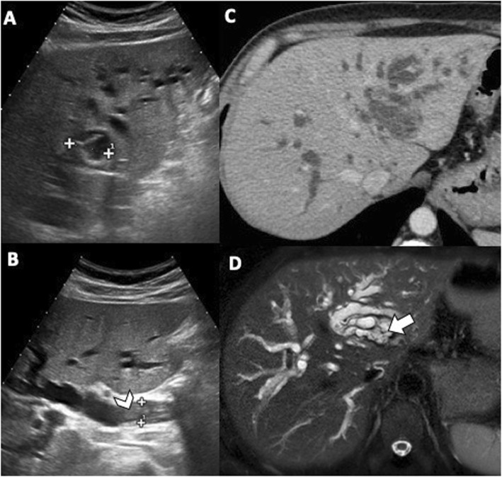
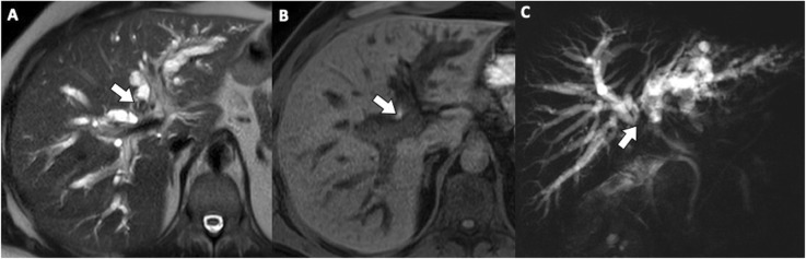
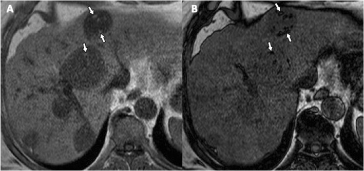
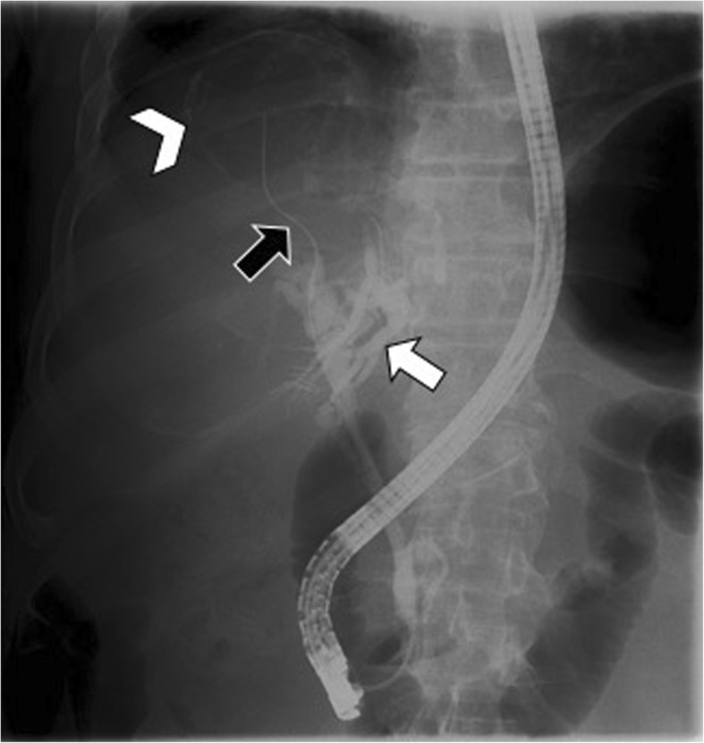
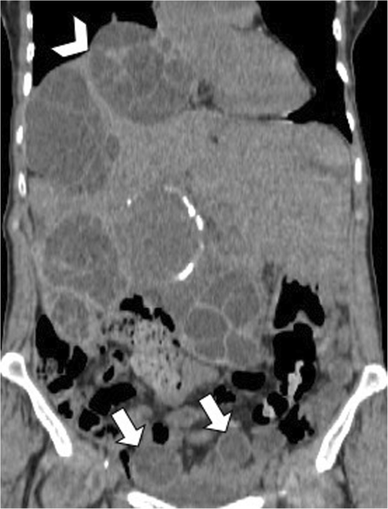
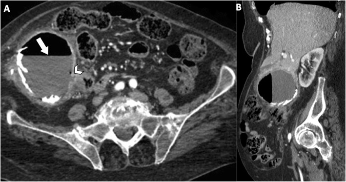
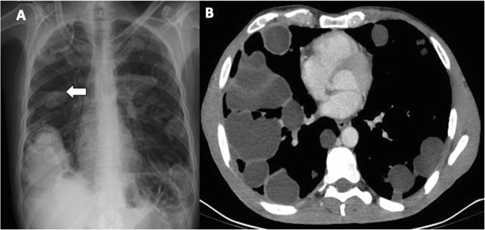
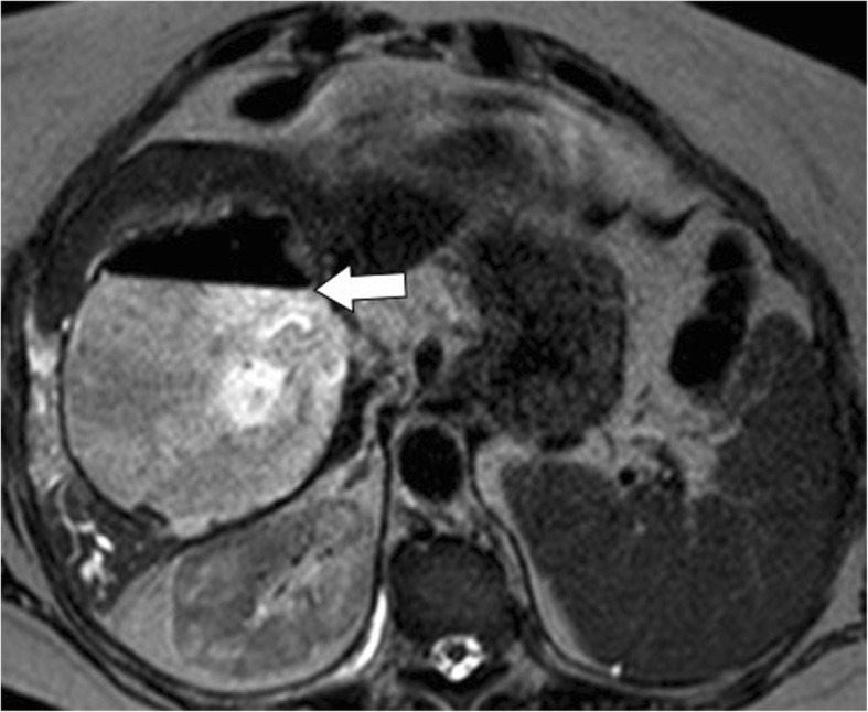
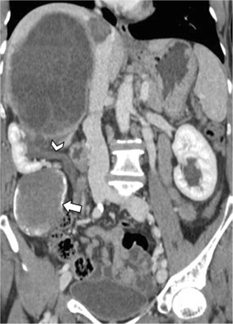
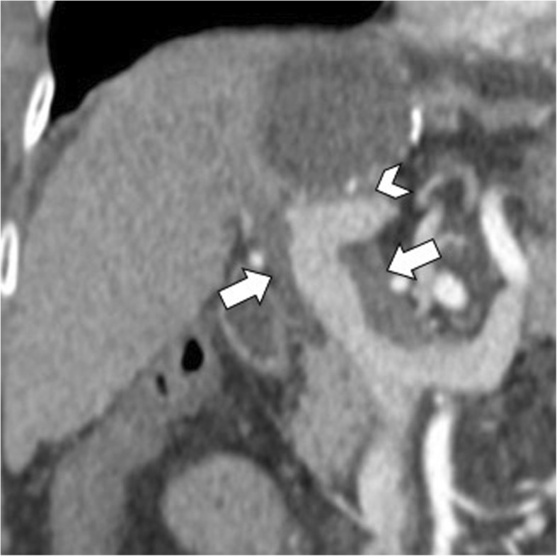
Hydatid disease is a worldwide zoonosis endemic in many countries. Liver echinococcosis accounts for 60-75% of cases and may be responsible for a wide spectrum of complications in about one third of patients. Some of these complications are potentially life-threatening and require prompt diagnosis and urgent intervention. In this article, we present our experience with common and uncommon complications of hepatic hydatid cysts which include rupture, bacterial superinfection, and mass effect-related complications. Specifically, the aim of this review is to provide key imaging features and diagnostic clues to guide the imaging diagnosis using a multimodality imaging approach, including ultrasound (US), computed tomography (CT), magnetic resonance (MR), and endoscopic retrograde cholangiopancreatography (ERCP).

Keywords: Computed tomography; Echinococcosis; Endoscopic retrograde cholangiopancreatography; Multimodal imaging.


Conflict of interest statement

Giuseppe Brancatelli has received lecture fees from Bayer; all the remaining authors declare they have no competing interests.

Figures

Fig. 1
Fig. 1
A 50-year-old woman with contained rupture hydatid cyst. Axial contrast-enhanced CT on portal venous phase shows hepatic hydatid cyst (arrow) with hyperattenuating serpentine linear membrane within the cyst (arrowhead)
Fig. 2
Fig. 2
A 65-year-old woman with contained rupture hydatid cyst due to response to therapy. Contrast-enhanced CT on portal venous phase shows the serpentine linear membrane within an almost completely calcified cyst
Fig. 3
Fig. 3
A 48-year-old man with hydatid liver cyst complicated with communicating rupture. a, b US images show a focal anechoic liver lesion with internal septa and hyperechoic peripheral rim, with some posterior shadows, diffuse biliary dilatation and endoluminal hyperechoic foci (arrowhead) into the common bile duct with no posterior acoustic shadows. Axial CT scan in the portal venous phase (c) and MR image on T2-weighted sequence (d) confirm the presence of a liver cyst and diffuse biliary dilatation, with material into the biliary tree (arrow). These features are consistent with progressive migration of the hydatid sand and membranes from the hilar confluence to the common bile duct
Fig. 4
Fig. 4
A 48-year-old man with communicating rupture. MR images on T2-weighted (a) and T1-weighted gradient echo (b) sequences demonstrate endoluminal biliary content (arrows), hypointense and hyperintense on respective images, at the hilar biliary confluence. On MRCP image (c), the endoluminal obstructive biliary content leads to loss of signal intensity (arrow) from the hilar biliary confluence to the common hepatic duct, while the common bile duct is patent
Fig. 5
Fig. 5
A 66-year-old man with multiple fat-containing hydatid cysts in the liver. a Axial gradient-echo in-phase T1-weighted MR image shows multiple high signal intensity foci (arrows) within hepatic hydatid cyst. b Axial gradient-echo opposed-phase T1-weighted MR image shows macroscopic fat within the same lesions, a finding confirmed by chemical shift artifact and signal cancelation (arrows) surrounding the lipid pure component
Fig. 6
Fig. 6
A 54-year-old man with air content within a hepatic hydatid cyst. Coronal reformatted CT image on portal venous phase shows air bubbles (arrowhead) within a unilocular calcified hydatid cyst. This finding was suggestive of occult cysto-biliary communication, which was later confirmed at surgery
Fig. 7
Fig. 7
A 65-year-old woman with cysto-biliary rupture. a Radiographic image performed during ERCP procedure shows multiple filling defects into the common bile duct (arrow), representing hydatid membranes. b Endoscopic image of the same patient shows hydatid membranes extraction
Fig. 8
Fig. 8
A 72-year-old man with communicating rupture. Radiographic image performed during ERCP demonstrates a linear calcified opacity (arrowhead)—representing a hepatic hydatid cyst—and intrahepatic bile duct dilatation (white arrow). The catheter (black arrow) introduced in biliary tree enters directly into the cyst, documenting the cysto-biliary fistula
Fig. 9
Fig. 9
A 66-year-old woman with diffuse abdominal echinococcosis. Coronal reformatted CT image shows peritoneal dissemination of hydatid cysts (arrows), due to direct rupture of the original liver cysts and compression of the right diaphragm by one of the hepatic hydatid cyst (arrowhead)
Fig. 10
Fig. 10
A 70-year-old man with echinococcosis. Axial CT image on portal venous phase demonstrates a pararectal homogeneous cystic lesion (arrow), showing internal septa and partial wall calcification, in a patient with hepatic hydatid cysts (not shown in this image)
Fig. 11
Fig. 11
An 85-year-old woman with hydatid cysto-colic fistulization. Axial contrast-enhanced CT image (a) and sagittal reformatted CT image (b) on portal venous phase demonstrate a large hepatic hydatid cyst with peripheral wall calcifications and air-fluid level (arrow), with frank communication (arrowhead) with the right colon
Fig. 12
Fig. 12
A 50-year-old man with hepatic and pleuro-pulmonary echinococcosis, secondary to trans-diaphragmatic dissemination. a Chest x-ray shows bilaterally multiple round cavitated lesions—some of which with air-fluid level (arrow)—as well as radiopaque lesions. b Chest CT scan of the same patient shows multiple cystic lesions, some of which appear stuffed with fluid-density material, others cavitated with air-fluid levels (not shown in this image)
Fig. 13
Fig. 13
A 66-year-old woman with infected hydatid cyst. Axial T2-weighted MR image demonstrates an air-fluid level (arrow) within a calcified hydatid cyst, but lack of frank cysto-biliary communication. This finding suggests superinfection of a ruptured hydatid cyst that was later proved at surgery
Fig. 14
Fig. 14
A 57-year-old man with communicating rupture and superinfection of liver hydatid cyst. Axial contrast-enhanced CT images at different levels (a, b) show multiple intrahepatic abscesses (arrowheads), adjacent to a hydatid cystic lesion with peripheral wall calcifications and intracystic air content. Note a calcified fragment of wall cyst within the right intrahepatic bile duct (white arrow), which migrates into the common bile duct (black arrow) at 10-days CT follow-up (c)
Fig. 15
Fig. 15
A 21-year-old man with hepatic hydatid cyst. Axial contrast-enhanced CT image on portal venous phase shows hepatic hydatid cyst (arrow) causing compression and dilatation of adjacent intrahepatic bile ducts (arrowhead)
Fig. 16
Fig. 16
A 60-year-old woman with large partially exophytic hepatic hydatid cyst. Coronal reformatted CT image on portal venous phase shows a large hepatic hydatid cyst with multiple daughter cysts, associated with peritoneal hydatid cysts (arrow). The exophytic growth of the hepatic hydatid cyst causes significant mass effect on the right kidney, which appears compressed and dislocated with hydronephrosis (arrowhead)
Fig. 17
Fig. 17
A 72-year-old woman with hydatid cyst and portal vein thrombosis. Oblique reformatted CT image on portal venous phase shows a large hepatic hydatid cyst adjacent to the left branch of portal vein (arrowhead) and partial portal vein thrombosis (arrows)

References

    1. https://www.who.int/echinococcosis/epidemiology/en/. Accessed 26 Jul 2019
    1. https://www.who.int/news-room/fact-sheets/detail/echinococcosis. Accessed 26 Jul 2019
    1. Eckert J, Gemmell MA, Meslin FX, Pawłowski ZS (2001) WHO/OIE manual on echinococcosis in humans and animals: a public health problem of global concern. Available via https://apps.who.int/iris/bitstream/handle/10665/42427/929044522X.pdf;js.... Accessed 26 Jul 2019
    1. Beggs I. The radiology of hydatid disease. American Journal of Roentgenology. 1985;145(3):639–648. doi: 10.2214/ajr.145.3.639. - DOI - PubMed
    1. Polat P, Kantarci M, Alper F, Suma S, Koruyucu MB, Okur A. Hydatid disease from head to toe. Radiographics. 2003;23:475–494. doi: 10.1148/rg.232025704. - DOI - PubMed

LinkOut - more resources