. 2020 Jan;24(2):1541-1552.
doi: 10.1111/jcmm.14839. Epub 2019 Dec 3.

PNPLA3 I148M mediates the regulatory effect of NF-kB on inflammation in PA-treated HepG2 cells

Affiliations

PNPLA3 I148M mediates the regulatory effect of NF-kB on inflammation in PA-treated HepG2 cells

Shuhua Yuan et al. J Cell Mol Med. 2020 Jan.

Abstract

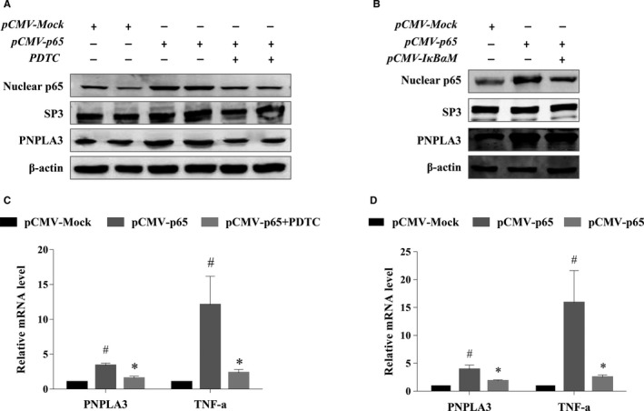
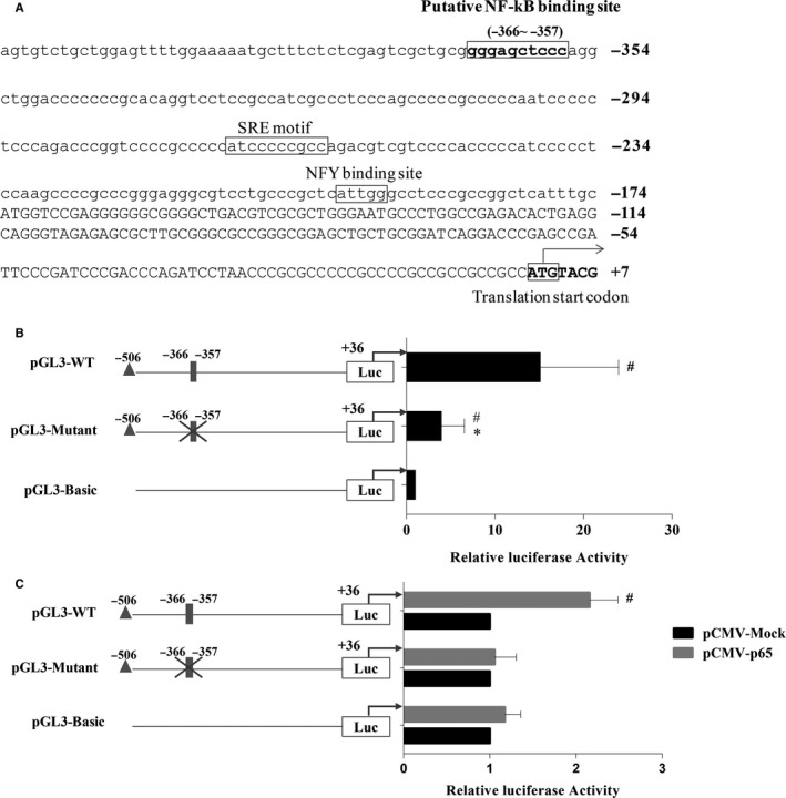
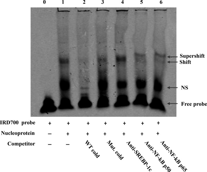
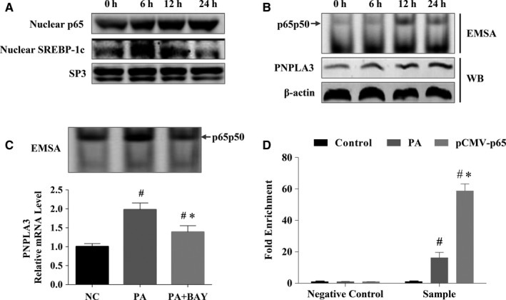
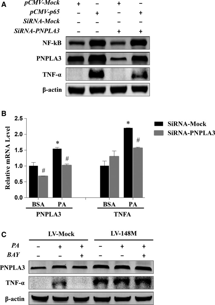
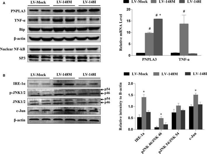
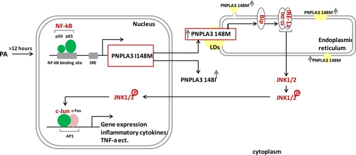
Both PNPLA3 I148M and hepatic inflammation are associated with nonalcoholic fatty liver disease (NAFLD) progression. This study aimed to elucidate whether PNPLA3 I148M is involved in NF-kB-related inflammation regulation in NAFLD. HepG2 cells homozygous for the PNPLA3 I148M mutation were used. The human PNPLA3 promoter sequence was screened for NF-kB binding sites using the MATCH and PATCH tools. NF-kB-mediated transcriptional regulation of the PNPLA3 gene was assessed by luciferase reporter assay, EMSA and ChIP-qPCR. Wild-type (I148I) and mutant (M148M) PNPLA3 were overexpressed using stable lentivirus-mediated transfection. The pCMV vector and siRNA were transiently transfected into cells to direct NF-kB overexpression and PNPLA3 silencing, respectively. A putative NF-kB binding site in the human PNPLA3 promoter was shown to be necessary for basal and NF-kB-driven transcriptional activation of PNPLA3 and protein/DNA complex formation. Supershift analysis demonstrated a protein/DNA complex specifically containing the NF-kB p65 and p50 subunits. ChIP-qPCR confirmed the endogenous binding of NF-kB to the human PNPLA3 promoter in response to NF-kB overexpression and palmitic acid (PA) challenge. The silencing of PNPLA3 blocked the overexpression of NF-kB or PA-induced TNF-α up-regulation. Moreover, mutant PNPLA3 overexpression prevented NF-kB inhibitor-induced down-regulation of TNF-α expression in PA-treated HepG2 cells. Finally, the overexpression of mutant but not wild-type PNPLA3 increased TNF-α expression and activated the ER stress-mediated and NF-kB-independent inflammatory IRE-1α/JNK/c-Jun pathway. Human PNPLA3 was shown to be a target of NF-kB, and PNPLA3 I148M mediated the regulatory effect of NF-kB on inflammation in PA-treated HepG2 cells, most likely via the IRE-1α/JNK/c-Jun ER stress pathway.

Keywords: ER stress; NAFLD; NF-kB; PNPLA3; inflammation.


Conflict of interest statement

The authors declare that they have no conflict of interest.

Figures

Figure 1
Figure 1
PNPLA3 expression was regulated by NF‐kB in HepG2 cells. HepG2 cells were transfected with blank pCMV (pCMV‐Mock) or pCMV‐p65 with or without pretreatment of NF‐kB inhibitor PDTC for 6 h, and then, the protein expression of PNPLA3 and nuclear NF‐kB (A), and mRNA expression of PNPLA3 and TNF‐α (C) were detected using Western blotting and real‐time PCR, respectively. HepG2 cells were transfected with pCMV‐Mock or pCMV‐p65 with or without pre‐transfection of pCMV‐IkBαM, and then, the protein expression of PNPLA3 and nuclear NF‐kB (B), and mRNA expression of PNPLA3 and TNF‐α (D) were detected using Western blotting and real‐time PCR, respectively. The results of real‐time PCR are presented as relative mRNA levels from three independent experiments normalized to the mock transfected control. #P < .05 compared with pCMV‐Mock, *P < .05 compared with pCMV‐p65
Figure 2
Figure 2
NF‐kB transactivated human PNPLA3 promoter through a putative NF‐kB binding site. A, Human PNPLA3 promoter upstream of the 5′UTR. The putative NF‐kB binding sites are highlighted with boxes; SREBP‐1c and NFY binding sites are underlined with a thick line. The ATG translation start codon where the A is numbered with 1 is indicated in bold and boxed. B, Relative luciferase activity of different PNPLA3 promoter‐reporter constructs. HepG2 cells were transfected with pGL3‐WT, PGL3‐Mutant and pGL3‐Basic reporter constructs for 24 h to measure the relative luciferase activities of PNPLA3 promoter by dual‐luciferase assays. Relative luciferase activity was corrected for Renilla luciferase activity and normalized to the pGL3‐Basic activity from three independent experiments. #P < .05 compared with pGL3‐Basic, *P < .05 compared with pGL3‐WT. C, NF‐kB‐driven relative luciferase activities from different PNPLA3 promoter‐reporter constructs in HepG2 cells. Each PNPLA3 promoter‐reporter construct was transiently cotransfected with pCMV‐Mock or pCMV‐p65 into HepG2 cells. Cell lysates were collected 24 h post‐cotransfection, and dual‐luciferase assays were performed. Relative luciferase activity was corrected for Renilla luciferase activity and normalized to the pCMV‐Mock activity from three independent experiments. # P < .05 compared with pCMV‐Mock. A grey‐filled rectangle represents the putative NF‐kB binding site, and a cross represents the mutation of the binding element
Figure 3
Figure 3
NF‐kB bound to the human PNPLA3 promoter. EMSA was performed using an IRD‐700‐labelled double‐stranded probe containing the putative NF‐kB binding sequence of human PNPLA3 gene promoter. The incubation of nuclear protein extracted from HepG2 cells and labelled probe was performed without or with the indicated unlabelled oligonucleotides (WT cold probe or Mut. cold probe) in the competition assays. And a supershift assay was performed to specifically assert the DNA‐protein interactions by using NF‐kB p65 and p50 antibody. SREBP‐1c antibody was used as control for supershift assay. Lane 1: no competitor; Lane 2: WT cold competitor (200×); Lane 3: mutant cold competitor (200×); Lane 4: SREBP‐1c antibody; Lane 5: NF‐kB p50 antibody; Lane 6: NF‐kB p56 antibody. The result is representative of two independent experiments with similar results
Figure 4
Figure 4
Binding of NF‐kB to human PNPLA3 promoter mediated expression of PNPLA3 during long‐term PA treatment. A, Nucleoprotein expression trait of NF‐kB and SREBP‐1c with time during PA treatment. Nucleoprotein levels of NF‐kB and SREBP‐1c were measured at 0 h and 6 h, 12 h, and 24 h post‐treatment of PA by Western blotting and normalized to SP3; B, PA increased nucleoproteins binding to PNPLA3 promoter containing the putative NF‐kB binding site. Nucleoproteins and cytoplasmic proteins were extracted from HepG2 cells at 0 h and 12 h and 24 h post‐treatment of PA. Nucleoproteins incubated with the IRD‐700‐labelled double‐stranded DNA (dsDNA) probe of human PNPLA3 promoter containing the putative NF‐kB binding site. The binding of protein and probe was detected by EMSA. The cytoplasmic PNPLA3 protein expression was detected by Western blotting; C, BAY‐117082 attenuated the binding of nucleoprotein extracted from HepG2 cells treated with PA for 24 h and probe of human PNPLA3 promoter containing the putative NF‐kB binding site. Nucleoproteins and mRNA were extracted from HepG2 cells 24 h post‐treatment of PA with or without pretreatment of BAY‐117082. The binding of nucleoproteins and the dsDNA probe of human PNPLA3 promoter containing the putative NF‐kB binding site was detected by EMSA. PNPLA3 mRNA level was detected by real‐time PCR. Results of real‐time PCR are presented as relative mRNA levels from three independent experiments normalized to the control. #P < .05 compared with NC, *P < .05 compared with PA D, PA increased the endogenous binding of NF‐kB and PNPLA3 promoter in vivo. The endogenous binding of NF‐kB and PNPLA3 promoter was detected by CHIP‐qPCR, which was performed using an anti‐NF‐KB P65 antibody and chromatin prepared from HepG2 treated with PA for 24 h or pCMV p65 for 24 h. Fold enrichment as percentage of input DNA was calculated. Rabbit IgG was used as a mock antibody for negative control in ChIP. #P < .05 compared with control; *P < .05 compared with PA
Figure 5
Figure 5
PNPLA3 mediated the NF‐kB regulation of inflammation in PA‐treated HepG2 cells. A, PNPLA3 mediated NF‐kB regulation of TNF‐α expression in HepG2 cells. HepG2 cells were transfected with pCMV‐p65, or cotransfected with pCMV‐p65 and siRNA‐PNPLA3. Cells transfected with the pCMV‐Mock or/and siRNA‐Mock were used as negative control. Cell lysates were collected 24 h post‐transfection. The protein expressions of NF‐kB, PNPLA3 and TNF‐α were detected by Western blotting. B, Inhibition of PNPLA3 alleviated PA‐induced hepatocyte inflammation. HepG2 cells were treated with PA for 24 h with pre‐transfection of siRNA‐Mock or siRNA‐PNPLA3. The mRNA levels of PNPLA3 and TNF‐α were detected by real‐time PCR. The results are presented as the mean ± SD from three independent experiments. #P < .05 compared with siRNA‐Mock‐transfected cells, *P < .05 compared with siRNA‐Mock‐transfected cells treated with BSA; C, Overexpression of PNPLA3 M148M prevented NF‐kB inhibitor‐induced reduction of PA‐related inflammation. HepG2 cells that stably transfected with LV‐148M or LV‐Mock were treated with PA for 24 h with or without pretreatment of BAY‐117082. Cell lysates were harvested to measure the protein expressions of PNPLA3 and TNF‐α by Western blotting
Figure 6
Figure 6
Overexpression of PNPLA3 M148M activated ER stress signal IRE‐1α‐JNK‐c‐Jun inflammatory pathway. A, PNPLA3 M148M overexpression increased TNF‐α expression in a NF‐kB independent way. HepG2 cells were stably infected with lentiviral PNPLA3 M148M (LV‐148M), lentiviral PNPLA3 I148I (LV‐148I) and mock lentivirus (LV‐Mock), respectively. Nucleoprotein and cytoplasmic protein were extracted after infection to measure protein expressions of nuclear NF‐kB, PNPLA3 and TNF‐α by Western blotting (left panel). RNA was extracted to measure PNPLA3 and TNF‐α mRNA levels by real‐time PCR (right panel). The real‐time PCR results are presented as the mean ± SD from three independent experiments. #P < .05 compared with LV‐Mock; *P < .05 compared with LV‐148M. B, PNPLA3 M148M but not I148I overexpression activated IRE‐1α‐JNK‐c‐Jun pathway. HepG2 cells were stably transfected with LV‐148M, LV‐148I and LV‐Mock, respectively. Cytoplasmic protein was extracted to measure protein expressions of IRE‐1α, total and phosphorylation JNK1/2, and c‐Jun by Western blotting (left panel). The relative band intensities (right panel) in Western blots (n = 2) were determined using ImageJ and normalized to β‐actin. Statistical significance was performed by one‐way ANOVA. Data are presented as the mean ± SD. *P < .05 compared with LV‐Mock
Figure 7
Figure 7
Scheme of PNPLA3 I148M‐related inflammatory signalling in HepG2 treated with PA. PNPLA3 gene is transcriptionally up‐regulated by NF‐kB during long‐term PA treatment. The distribution of wild‐type and mutant PNPLA3 proteins is different: I148M mutant proteins are distributed in lipid droplets, while wild‐type proteins are distributed in the cytoplasm. PNPLA3 I148M protein then activates the IRE1a signalling of ER stress, followed by phosphorylating JNK1/2 and up‐regulating c‐Jun expression, which finally up‐regulates c‐Jun‐dependent expression of inflammatory cytokines, such as TNF‐α

Similar articles

Cited by

References

    1. Zeng MD, Fan JG, Lu LG, et al. Chinese national consensus workshop on nonalcoholic fatty liver disease. Guidelines for the diagnosis and treatment of nonalcoholic fatty liver diseases. J Dig Dis. 2008;9:108‐112. - PubMed
    1. Lewis JR, Mohanty SR. Nonalcoholic fatty liver disease: a review and update. Dig Dis Sci. 2010;55:560‐578. - PubMed
    1. Mishra P, Younossi ZM. Current treatment strategies for non‐alcoholic fatty liver disease (NAFLD). Curr Drug Discov Technol. 2007;4:133‐140. - PubMed
    1. Romeo S, Kozlitina J, Xing C, et al. Genetic variation in PNPLA3 confers susceptibility to nonalcoholic fatty liver disease. Nat Genet. 2008;40:1461‐1465. - PMC - PubMed
    1. Pingitore P, Pirazzi C, Mancina RM, et al. Recombinant PNPLA3 protein shows triglyceride hydrolase activity and its I148M mutation results in loss of function. Biochim Biophys Acta. 2014;1841:574‐580. - PubMed

Publication types

MeSH terms