. 2019 Dec 1;75(Pt 12):1040-1050.
doi: 10.1107/S2059798319010179. Epub 2019 Nov 19.

Non-merohedral twinning: from minerals to proteins

Affiliations

Non-merohedral twinning: from minerals to proteins

Madhumati Sevvana et al. Acta Crystallogr D Struct Biol. .

Abstract

In contrast to twinning by merohedry, the reciprocal lattices of the different domains of non-merohedral twins do not overlap exactly. This leads to three kinds of reflections: reflections with no overlap, reflections with an exact overlap and reflections with a partial overlap of a reflection from a second domain. This complicates the unit-cell determination, indexing, data integration and scaling of X-ray diffraction data. However, with hindsight it is possible to detwin the data because there are reflections that are not affected by the twinning. In this article, the successful solution and refinement of one mineral, one organometallic and two protein non-merohedral twins using a common strategy are described. The unit-cell constants and the orientation matrices were determined by the program CELL_NOW. The data were then integrated with SAINT. TWINABS was used for scaling, empirical absorption corrections and the generation of two different data files, one with detwinned data for structure solution and refinement and a second one for (usually more accurate) structure refinement against total integrated intensities. The structures were solved by experimental phasing using SHELXT for the first two structures and SHELXC/D/E for the two protein structures; all models were refined with SHELXL.

Keywords: non-merohedral twinning; twinned structure refinement; twinned structure solution.


Figures

Figure 1
Figure 1
Diffraction patterns in APEX (Bruker’s crystallography software suite; Bruker, 2018 ▸) (a) indicating unindexed reflections (black arrow) and split reflections (white arrow) and (b) showing a split reflection profile
Figure 2
Figure 2
Reciprocal-space plot of the k = 2 layer of a monoclinic structure (a) and the overlay of this plot with a rotated plot simulating non-merohedral twinning (b).
Figure 3
Figure 3
Schematic picture of reflections from two domains (blue and red) with different degrees of overlap. The rectangles represent the integration boxes. (a) Only one orientation matrix is used; (b) both orientation matrices are used.
Figure 4
Figure 4
Structure of chromite with the Fe2+ tetrahedron in orange and the Cr3+ octahedron in blue, produced with VESTA v.3.4.6 (Momma & Izumi, 2011 ▸).
Figure 5
Figure 5
RLATT plot showing both orientations for chromite.
Figure 6
Figure 6
RLATT plot showing both orientations for Cp*2MeZrOTiMe2Cp*.
Figure 7
Figure 7
Structure of one of the two molecules of Cp*2MeZrOTiMe2Cp*.
Figure 8
Figure 8
RLATT plot showing the two orientations of insulin.
Figure 9
Figure 9
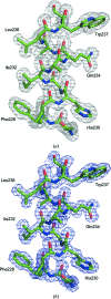
Part of the SHELXE map (a) and the final refined map (b) for cubic insulin contoured at 1σ.
Figure 10
Figure 10
An example image of a glucose isomerase triplet. (a) An image taken at 2θ = 40° and a detector distance of 18 cm. (b) The indexed image using CELL_NOW. The first domain is coloured blue, the second domain is in green and the third domain is in red.
Figure 11
Figure 11
Normalized scale factor against run/frame number from TWINABS for (a) cubic insulin and (b) glucose isomerase; domain 1 is coloured blue, domain 2 is in red and domain 3 (only for the triple twin of glucose isomerase) is in green. The corresponding crystal pictures demonstrate the correlation between crystal growth and different centring in the beam.
Figure 12
Figure 12
Part of the SHELXE map (a) and the final refined map (b) for glucose isomerase contoured at 1σ.

References

    1. Adams, P. D., Afonine, P. V., Bunkóczi, G., Chen, V. B., Davis, I. W., Echols, N., Headd, J. J., Hung, L.-W., Kapral, G. J., Grosse-Kunstleve, R. W., McCoy, A. J., Moriarty, N. W., Oeffner, R., Read, R. J., Richardson, D. C., Richardson, J. S., Terwilliger, T. C. & Zwart, P. H. (2010). Acta Cryst. D66, 213–221. - PMC - PubMed
    1. Banumathi, S., Zwart, P. H., Ramagopal, U. A., Dauter, M. & Dauter, Z. (2004). Acta Cryst. D60, 1085–1093. - PubMed
    1. Battye, T. G. G., Kontogiannis, L., Johnson, O., Powell, H. R. & Leslie, A. G. W. (2011). Acta Cryst. D67, 271–281. - PMC - PubMed
    1. Betteridge, P. W., Carruthers, J. R., Cooper, R. I., Prout, K. & Watkin, D. J. (2003). J. Appl. Cryst. 36, 1487.
    1. Bourhis, L. J., Dolomanov, O. V., Gildea, R. J., Howard, J. A. K. & Puschmann, H. (2015). Acta Cryst. A71, 59–75. - PMC - PubMed

MeSH terms