Review
. 2020 Feb:60:12-20.
doi: 10.1016/j.conb.2019.11.005. Epub 2019 Nov 30.

Egocentric and allocentric representations of space in the rodent brain

Affiliations
Review

Egocentric and allocentric representations of space in the rodent brain

Cheng Wang et al. Curr Opin Neurobiol. 2020 Feb.

Abstract

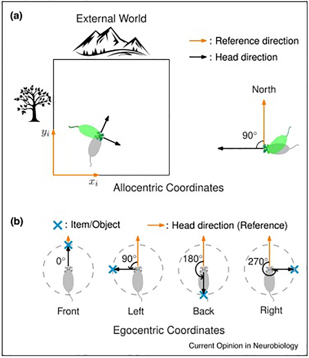
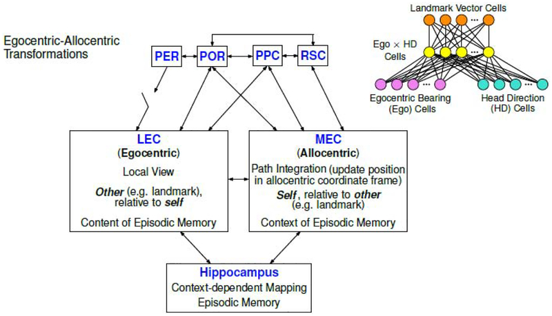
Spatial signals are prevalent within the hippocampus and its neighboring regions. It is generally accepted that these signals are defined with respect to the external world (i.e., a world-centered, or allocentric, frame of reference). Recently, evidence of egocentric processing (i.e., self-centered, defined relative to the subject) in the extended hippocampal system has accumulated. These results support the idea that egocentric sensory information, derived from primary sensory cortical areas, may be transformed to allocentric representations that interact with the allocentric hippocampal system. We propose a framework to explain the implications of the egocentric-allocentric transformations to the functions of the medial temporal lobe memory system.


Conflict of interest statement

Declaration of interests

The authors declare that they have no known competing financial interests or personal relationships that could have appeared to influence the work reported in this paper.

Figures

Figure 1.
Figure 1.. Definition of frames of reference for allocentric and egocentric coding.
(a) Representation of location and bearing in an allocentric reference frame. Left, in allocentric coordinates, the position of the subject is not dependent on the animal’s body or head orientation. Right, the allocentric head direction representation of the animal is defined with respect to an external reference direction, e.g. North. The allocentric head direction is independent of the body orientation. (b) Representation of the distance and bearing of external items in an egocentric reference frame. The egocentric representation of an external item/object is defined with respect to the animal itself. For a given item/object, a vector from the head of the subject to the item is constructed. The egocentric bearing of the item is defined as the angle of the vector, referenced to the subject’s allocentric head direction. Reproduced from Wang, Chen et al. [33].
Figure 2.
Figure 2.. Egocentric and allocentric representations outside the hippocampus
(a) Activity of some head direction cells in the postsubiculum were modulated by the egocentric bearing of the environmental boundary. In this example cell, the firing rate is much higher when the wall is on the animal’s left than when the wall is on its right. Modified, with permission, from Peyrache et al. [25]. (b) An example cell in postrhinal cortex showed strong activity in response to the presence of a border at a given egocentric direction and distance to the rat. The rat is in the center of this egocentric boundary rate map, in which distance and egocentric direction to the boundaries is indicated by radial distance and angle from the center, respectively, of this map. The yellow area indicates that the cell was most active when there was a border 10 cm to the left of the rat. Reproduced, with permission, from Gofman et al. [26]. (c) Parietal cortex is tuned to both allocentric and egocentric reference frames. Left, Schematic of egocentric cue direction (ECD, inner circle, numbers in blue) and head direction (outer circle, numbers in red). In this frame, the ECD is approximately 10° and head direction is 160°. Middle, a cell showed selectivity for ECD. Right, a cell showed conjunctive selectivity for ECD and head direction. Reproduced, with permission, from Wilber et al. [16]. (d) A substantial portion of cells in the dorsomedial striatum respond to environmental boundaries in an egocentric reference frame. Left, Trajectories of the rat (gray) superimposed with locations of the rat when the cell fires spikes (colored dots). Middle, Color wheel indicates the movement direction of the rat for each colored dot in the left panel. Right, Egocentric boundary rate map similar to that shown in (b). For this cell, the preferred orientation of the boundary is −98° (the boundary is to the left of the rat) and the preferred distance is approximately 5.5 cm from the boundary. Reproduced, with permission, from Hinman et al. [34]. (e) Some cells in POR encode the egocentric bearing of the center of the arena. Left, same as in (d), except that the color bar indicates the head direction instead of movement direction. Middle, Schematic of egocentric bearing of the center of the arena (“center-bearing”). Right, this example cell has the highest firing activity when the center bearing is 180° (i.e., behind the rat). Reproduced, with permission, from LaChance et al. [27].
Figure 3.
Figure 3.. Egocentric representation in the hippocampus and lateral entorhinal cortex
(a) Some hippocampal CA1 cells in bats showed egocentric goal direction selectivity. Left, Schematic for egocentric goal direction. Top right, an example goal direction cell that has the highest firing rate when the goal direction is 0° (the bat flies toward the goal location). Bottom right, trajectories of goal-direction angles along the behavioral session (gray), with spikes overlaid (red). Reproduced, with permission, from Sarel, Finkelstein et al. [28]. (b) Activity of some place cells in mouse hippocampal CA1 can be modulated by egocentric heading direction to a reference point in the environment. Left, Schematic of the egocentric heading direction relative to a specific reference point. Right, Heat map: spatial firing rate map; red circle: center of mass for the rate map; blue arrows: heading direction tuning within each spatial bin; black circle: the reference point obtained by a model based on the real heading direction tuning; red arrows: heading direction tuning fitted by the model in each spatial bin. Reproduced, with permission, from Jercog et al. [29]. (c) An example cell in LEC showed selectivity for egocentric bearing of the arena boundary/center. Left, trajectory (gray lines) and position and head direction of the rat when the cell fired (colored dots). Middle, Color wheel denotes the head direction. Right, Local head direction tuning in each spatial bin. Arrow direction: preferred head direction; arrow size: firing rate; color saturation: the mean vector length of the tuning curve (MVL); number on top: maximum MVL. (d) An example LEC cell tuned for egocentric bearing of goal location in a goal-oriented task. In this task, a single food well (red circle) was shifted from the standard goal location in session 1 (left) to a different location in session 2 (middle), and then back to the original standard location in session 3 (right). Local head direction tuning is showed as in (c). (e) An example cell in MEC showed selectivity for allocentric head direction, as demonstrated by all arrows pointing in the same, allocentric direction. (f) LEC represents spatial information in an egocentric frame of reference, whereas MEC utilizes an allocentric frame of reference. Bayesian information criterion (BICBoundary) indicates the goodness of fit of a cell’s activity to an egocentric bearing model or an allocentric bearing model. ΔBICBoundary describes the difference of goodness of fit between the two models, in which a negative value means the cell prefers the egocentric frame of reference, whereas a positive value means the cell prefers the allocentric frame of reference. (c-f) Reproduced, with permission, from Wang, Chen et al. [33].’
Figure 4.
Figure 4.. Working model for the egocentric-allocentric transformation in the medial temporal lobe and related brain regions.
In this highly simplified model of functional anatomy, perirhinal cortex (PER) and postrhinal cortex (POR) are two important parahippocampal regions that directly project to the entorhinal cortex (EC). (Note that this diagram dos not attempt to depict the relative strength of any of the connections.) The PER input is “gated” by the necessity for coactive inputs from other regions that presumably convey some type of salience tag (e.g., from amygdala or prefrontal cortex) to the PER sensory input [37]. Two high-order associative brain areas, posterior parietal cortex (PPC) and retrosplenial cortex (RSC), also have connections with the EC. Cells representing egocentric and allocentric reference frames were found recently in these brain regions upstream of the EC-hippocampus system. Upper right, a diagram (modified, with permission, from McNaughton et al. [15]) showing how specific circuits wired by cells encoding egocentric bearing and allocentric head direction information in these areas could construct an allocentric spatial signal (in this case, a landmark vector cell). Lateral entorhinal cortex (LEC) and medial entorhinal cortex (MEC), the two major cortical input regions to the hippocampus, show a functional dichotomy in egocentric vs. allocentric spatial frames of reference [33]. This difference is hypothesized to underlie separate roles of these regions in providing information to the hippocampal episodic memory system.

References

    1. O’Keefe J, Nadel L: The Hippocampus as a Cognitive Map. Oxford: Clarendon Press; 1978.
    1. Taube JS, Muller RU, Ranck JB: Head-direction cells recorded from the postsubiculum in freely moving rats. I. Description and quantitative analysis. J Neurosci 1990, 10:420–435. - PMC - PubMed
    1. Hafting T, Fyhn M, Molden S, Moser M-B, Moser EI: Microstructure of a spatial map in the entorhinal cortex. Nature 2005, 436:801–806. - PubMed
    1. McNaughton BL, Leonard B, Chen L: Cortical-hippocampal interactions and cognitive mapping: A hypothesis based on reintegration of the parietal and inferotemporal pathways for visual processing. Psychobiology 1989, 17:230–235.
    1. Navratilova Z, Hoang LT, Schwindel CD, Tatsuno M, McNaughton BL: Experience-dependent firing rate remapping generates directional selectivity in hippocampal place cells. Front Neural Circuits 2012, 6. - PMC - PubMed

Publication types