

Disrupted functional and structural connectivity within default mode network contribute to WMH-related cognitive impairment
- PMID: 31795048
- PMCID: PMC6861557
- DOI: 10.1016/j.nicl.2019.102088
Disrupted functional and structural connectivity within default mode network contribute to WMH-related cognitive impairment
Abstract
Aims: The prevalence of white matter hyperintensities (WMH) rises dramatically with aging. Both the progression of WMH and changing patterns of default mode network (DMN) have been proven to be closely associated with cognitive function. The present study hypothesized that changes in functional connectivity and structural connectivity of DMN contributed to WMH related cognitive impairment.
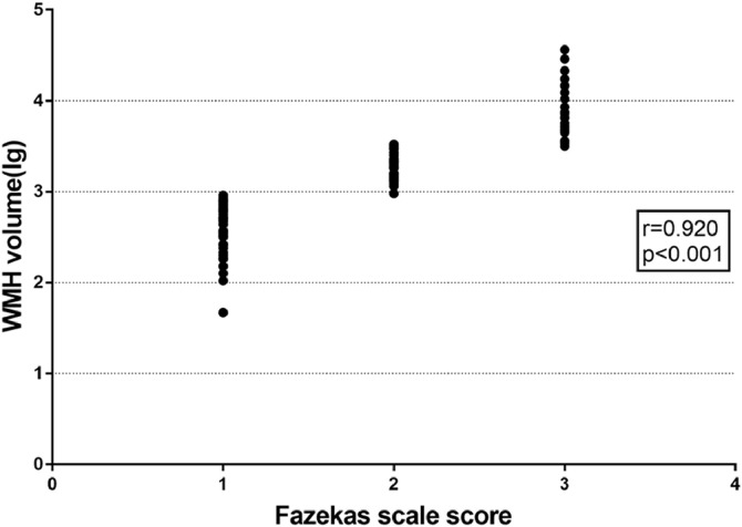
Methods: A total of 116 subjects were enrolled from the Cerebral Small Vessel Disease Register in Drum Tower Hospital of Nanjing University, and were distributed across three categories according to Fazekas rating scale: WMH I (n = 57), WMH II (n = 34), and WMH III(n = 25). All participants underwent neuropsychological tests and multimodal MRI scans, including diffusion tensor imaging and resting-state fMRI imaging. The alterations of functional connectivity and structural connectivity within the DMN were further explored.
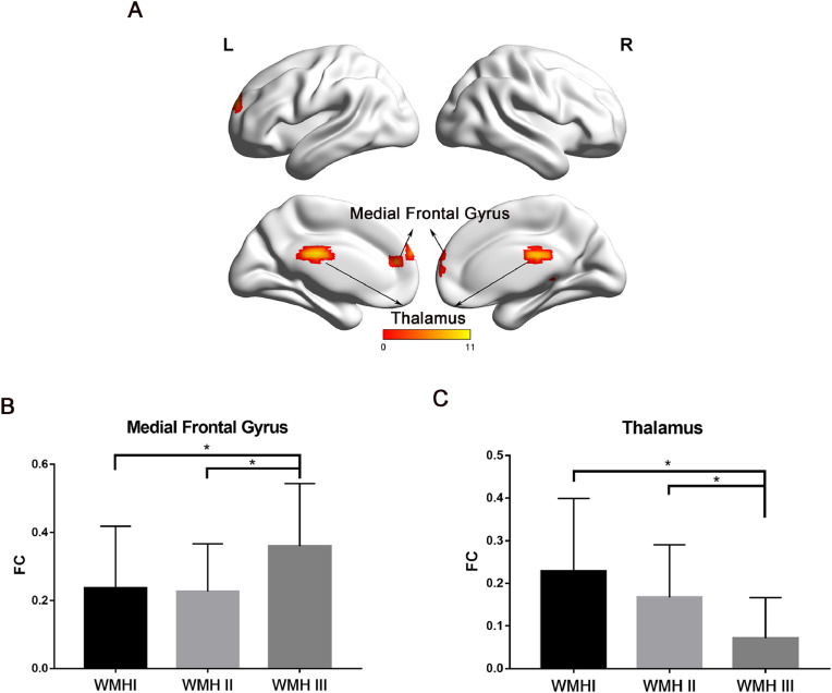
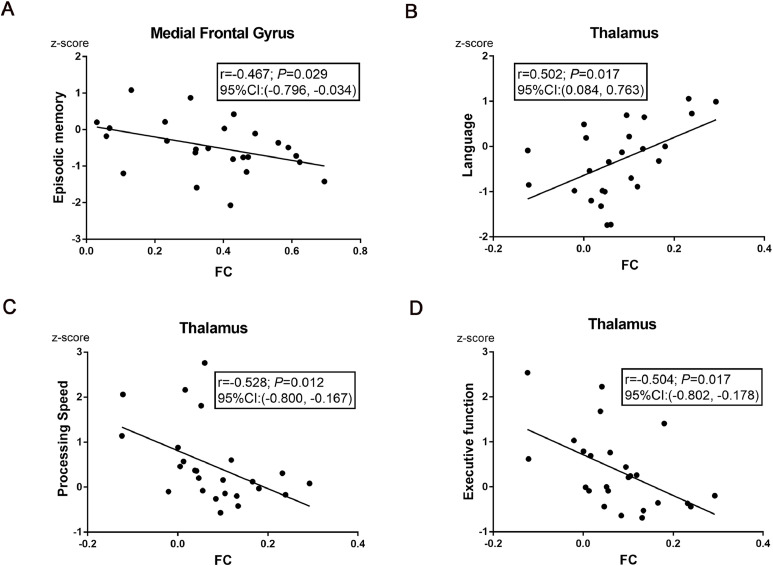
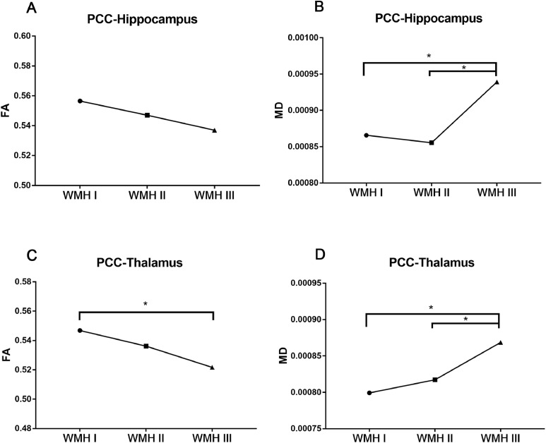
Results: Age and hypertension were risk factors for WMH progression. Subjects with a higher WMH burden displayed higher DMN functional connectivity in the medial frontal gyrus, while lower DMN functional connectivity in the thalamus. After adjusting for aging, gender, and education, the increased DMN functional connectivity in the medial frontal gyrus, and the increased mean diffusivity of the white matter tracts between the hippocampus and posterior cingulate cortex were independent indicators of worse performance in memory. Moreover, the decreased DMN functional connectivity in the thalamus and increased mean diffusivity of the white matter tracts between the thalamus and posterior cingulate cortex were independent risk factors for a slower processing speed.
Conclusion: The changes in functional connectivity and structural connectivity within the DMN attributed to WMH progression were responsible for the development of cognitive impairment.
Keywords: Cognitive impairment; Default mode network; Functional connectivity; Structural connectivity; White matter hyperintensities.
Copyright © 2019 The Author(s). Published by Elsevier Inc. All rights reserved.
Conflict of interest statement
The authors declare no conflict of interest with respect to the research, authorship, and/or publication of this article.
Figures
References
-
- Alber J., Alladi S., Bae H.J., Barton D.A., Beckett L.A., Bell J.M., Berman S.E., Biessels G.J., Black S.E., Bos I., Bowman G.L., Brai E., Brickman A.M., Callahan B.L., Corriveau R.A., Fossati S., Gottesman R.F., Gustafson D.R., Hachinski V., Hayden K.M., Helman A.M., Hughes T.M., Isaacs J.D., Jefferson A.L., Johnson S.C., Kapasi A., Kern S., Kwon J.C., Kukolja J., Lee A., Lockhart S.N., Murray A., Osborn K.E., Power M.C., Price B.R., Rhodius-Meester H.F.M., Rondeau J.A., Rosen A.C., Rosene D.L., Schneider J.A., Scholtzova H., Shaaban C.E., Silva N., Snyder H.M., Swardfager W., Troen A.M., van Veluw S.J., Vemuri P., Wallin A., Wellington C., Wilcock D.M., Xie S.X., Hainsworth A.H. White matter hyperintensities in vascular contributions to cognitive impairment and dementia (VCID): knowledge gaps and opportunities. Alzheimers Dement (N Y) 2019;5:107–117. - PMC - PubMed
-
- Alexander G.E., Chen K., Pietrini P., Rapoport S.I., Reiman E.M. Longitudinal pet evaluation of cerebral metabolic decline in dementia: a potential outcome measure in alzheimer’s disease treatment studies. Am. J. Psychiatry. 2002;159:738–745. - PubMed
-
- Bergsland N., Zivadinov R., Dwyer M.G., Weinstock-Guttman B., Benedict R.H. Localized atrophy of the thalamus and slowed cognitive processing speed in MS patients. Mult. Scler. 2016;22:1327–1336. - PubMed
-
- Bisecco A., Stamenova S., Caiazzo G., d'Ambrosio A., Sacco R., Docimo R., Esposito S., Cirillo M., Esposito F., Bonavita S., Tedeschi G., Gallo A. Attention and processing speed performance in multiple sclerosis is mostly related to thalamic volume. Brain Imaging Behav. 2018;12:20–28. - PubMed
Publication types
MeSH terms
LinkOut - more resources
Full Text Sources
Medical
