

Connectivity strength, time lag structure and the epilepsy network in resting-state fMRI
- PMID: 31795065
- PMCID: PMC6881607
- DOI: 10.1016/j.nicl.2019.102035
Connectivity strength, time lag structure and the epilepsy network in resting-state fMRI
Abstract
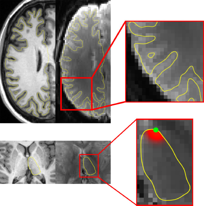
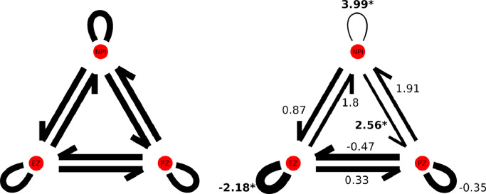
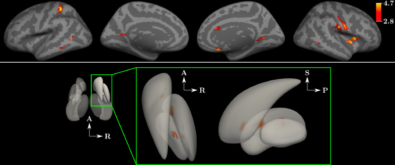
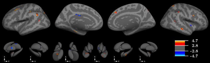
The relationship between the epilepsy network, intrinsic brain networks and hypersynchrony in epilepsy remains incompletely understood. To converge upon a synthesized understanding of these features, we studied two elements of functional connectivity in epilepsy: correlation and time lag structure using resting state fMRI data from both SEEG-defined epileptic brain regions and whole-brain fMRI analysis. Functional connectivity (FC) was analyzed in 15 patients with epilepsy and 36 controls. Correlation strength and time lag were selected to investigate the magnitude of and temporal interdependency across brain regions. Zone-based analysis was carried out investigating directed correlation strength and time lag between both SEEG-defined nodes of the epilepsy network and between the epileptogenic zone and all other brain regions. Findings were compared between patients and controls and against a functional atlas. FC analysis on the nodal and whole brain levels identifies consistent patterns of altered correlation strength and altered time lag architecture in epilepsy patients compared to controls. These patterns include 1) broadly distributed increased strength of correlation between the seizure onset node and the remainder of the brain, 2) decreased time lag within the seizure onset node, and 3) globally increased time lag throughout all regions of the brain not involved in seizure onset or propagation. Comparing the topographic distribution of findings against a functional atlas, all resting state networks were involved to a variable degree. These local and whole brain findings presented here lead us to propose the network steal hypothesis as a possible mechanistic explanation for the non-seizure clinical manifestations of epilepsy.
Keywords: Epilepsy network; Functional connectivity; Resting state networks.
Copyright © 2019 The Authors. Published by Elsevier Inc. All rights reserved.
Figures
References
-
- Bartolomei F., Bettus G., Stam C.J., Guye M. Interictal network properties in mesial temporal lobe epilepsy: a graph theoretical study from intracerebral recordings. Clin. Neurophysiol. 2013;124(12):2345–2353. https://www.ncbi.nlm.nih.gov/pubmed/23810635 Retrieved from. - PubMed
-
- Bartolomei F., Lagarde S., Wendling F., McGonigal A., Jirsa V., Guye M., Benar C. Defining epileptogenic networks: contribution of SEEG and signal analysis. Epilepsia. 2017 https://www.ncbi.nlm.nih.gov/pubmed/28543030 Retrieved from. - PubMed
-
- Bartolomei F., Wendling F., Bellanger J.J., Regis J., Chauvel P. Neural networks involving the medial temporal structures in temporal lobe epilepsy. Clin. Neurophysiol. 2001;112(9):1746–1760. https://www.ncbi.nlm.nih.gov/pubmed/11514258 Retrieved from. - PubMed
-
- Bartolomei F., Wendling F., Regis J., Gavaret M., Guye M., Chauvel P. Pre-ictal synchronicity in limbic networks of mesial temporal lobe epilepsy. Epilepsy Res. 2004;61(1–3):89–104. https://www.ncbi.nlm.nih.gov/pubmed/15451011 Retrieved from. - PubMed
-
- Besson P., Bandt S.K., Proix T., Lagarde S., Jirsa V.K., Ranjeva J.P., Guye M. Anatomic consistencies across epilepsies: a stereotactic-EEG informed high-resolution structural connectivity study. Brain. 2017;140(10):2639–2652. https://www.ncbi.nlm.nih.gov/pubmed/28969369 Retrieved from. - PubMed
