

Medical infrared thermal imaging of canine appendicular bone neoplasia
- PMID: 31796069
- PMCID: PMC6889724
- DOI: 10.1186/s12917-019-2180-6
Medical infrared thermal imaging of canine appendicular bone neoplasia
Abstract
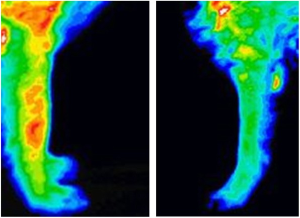
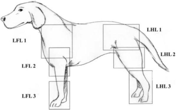
Background: Medical infrared thermal imaging (MITI) is a noninvasive imaging modality used in veterinary medicine as a screening tool for musculoskeletal and neurological disease processes. An infrared camera measures the surface body heat and produces a color map that represents the heat distribution. Local trauma or disease can impair the autonomic nervous system, which leads to changes in the local dermal microcirculation and subsequent alteration of surface body heat. Disruption of autonomic flow to the cutaneous vasculature at deeper levels can also result in asymmetric thermographic results. The purpose of this study was to evaluate surface temperature differences between limbs affected by bone neoplasia and their normal contralateral limbs.
Results: A statistically significant difference in average temperature was noted between regions of interest of the two groups (paired difference: 0.53 C° ± 0.14; P = 0.0005). In addition, pattern recognition analysis yielded a 75-100% success rate in lesion identification.
Conclusions: Significant alterations noted with average temperature and thermographic patterns indicate that MITI can document discernible changes associated with the presence of canine appendicular bone tumors. While MITI cannot be used as the sole diagnostic tool for bone cancer, it can be used as a screening modality and may be applicable in early detection of cancer.
Keywords: Canine appendicular bone cancer; Medical infrared thermal imaging.
Conflict of interest statement
The authors declare that they have no competing interests.
Figures
References
-
- Tjalma RA. Canine bone sarcoma: estimation of relative risk as a function of body size. J Natl Cancer Inst. 1966;36:1137–1150. - PubMed
-
- Kudnig ST, Séguin B. Veterinary surgical oncology. 1. West Sussex: Wiley; 2012. Musculoskeletal system; pp. 491–568.
-
- Tobias KM, Johnston SA. Veterinary surgery small animal. 2. St. Louis: Elsevier; 2018. Musculoskeletal Neoplasia and limb-sparing surgery; pp. 1347–1372.
-
- Liptak JM, Dernell WS, Ehrhart NP. Canine appendicular osteosarcoma: diagnosis and palliative treatment. Compendium. 2004;26:172–183.
MeSH terms
LinkOut - more resources
Full Text Sources
Medical
