Review
. 2019 Dec 3;14(1):279.
doi: 10.1186/s13023-019-1260-9.

Developmental hypomyelination in Wolfram syndrome: new insights from neuroimaging and gene expression analyses

Affiliations
Review

Developmental hypomyelination in Wolfram syndrome: new insights from neuroimaging and gene expression analyses

Amjad Samara et al. Orphanet J Rare Dis. .

Abstract

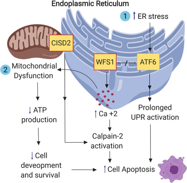
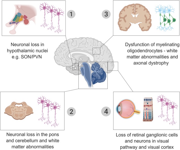
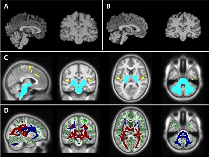
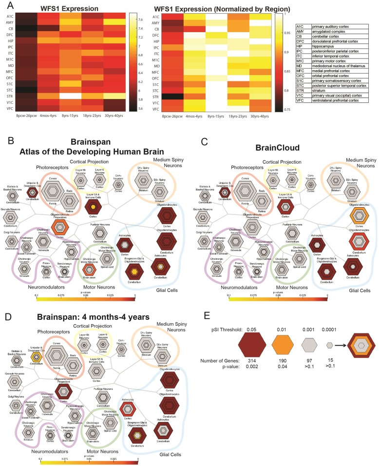
Wolfram syndrome is a rare multisystem disorder caused by mutations in WFS1 or CISD2 genes leading to brain structural abnormalities and neurological symptoms. These abnormalities appear in early stages of the disease. The pathogenesis of Wolfram syndrome involves abnormalities in the endoplasmic reticulum (ER) and mitochondrial dynamics, which are common features in several other neurodegenerative disorders. Mutations in WFS1 are responsible for the majority of Wolfram syndrome cases. WFS1 encodes for an endoplasmic reticulum (ER) protein, wolframin. It is proposed that wolframin deficiency triggers the unfolded protein response (UPR) pathway resulting in an increased ER stress-mediated neuronal loss. Recent neuroimaging studies showed marked alteration in early brain development, primarily characterized by abnormal white matter myelination. Interestingly, ER stress and the UPR pathway are implicated in the pathogenesis of some inherited myelin disorders like Pelizaeus-Merzbacher disease, and Vanishing White Matter disease. In addition, exploratory gene-expression network-based analyses suggest that WFS1 expression occurs preferentially in oligodendrocytes during early brain development. Therefore, we propose that Wolfram syndrome could belong to a category of neurodevelopmental disorders characterized by ER stress-mediated myelination impairment. Further studies of myelination and oligodendrocyte function in Wolfram syndrome could provide new insights into the underlying mechanisms of the Wolfram syndrome-associated brain changes and identify potential connections between neurodevelopmental disorders and neurodegeneration.

Keywords: Hypomyelination; Neurodegeneration; Neurodevelopment; Neuroimaging; Unfolded protein response; WFS1, endoplasmic reticulum stress.


Conflict of interest statement

The authors declare that they have no competing interests..

Figures

Fig. 1
Fig. 1
A schematic representation of the endoplasmic reticulum and mitochondrial molecular changes in Wolfram syndrome (the red box indicate a deficiency of this protein). ER: endoplasmic reticulum; ATF6: Activating transcription factor 6; UPR: unfolded protein response; WFS1: wolframin protein; CISD2: CISD2 protein product, ERIS
Fig. 2
Fig. 2
Brain structures and tissues most prominently affected in Wolfram syndrome. SON: supraoptic nucleus; PVN: paraventricular nucleus
Fig. 3
Fig. 3
a) Sagittal and coronal view of a healthy young adult brain. b) Sagittal and coronal view of a young adult brain with Wolfram syndrome. c) Significant volumetric differences between Wolfram syndrome and controls, controlling for whole brain volume. Regions that are smaller in Wolfram syndrome are in light-blue, while regions that are larger are in yellow. d) White matter microstructure alterations in Wolfram syndrome as measured by diffusion tensor imaging. Green: white matter skeleton created by tract-based spatial statistics skeletonization step; Blue: white matter tracts with greater radial diffusivity in Wolfram syndrome; Yellow: lower fractional anisotropy; Red: white matter tracts with overlap of greater radial diffusivity and lower fractional anisotropy is shown in red
Fig. 4
Fig. 4
Temporal expression of WFS1 and cell type-specific expression of WFS1-related genes. a) Left: Mean WFS1 spatiotemporal expression (RPKM, or reads per kilobase per million) in 16 brain regions and 5 developmental time periods from the BrainSpan database (8–26 post-conception weeks (pcw), 4 months-4 years, 8 years–15 years, 18 years–23 years, and 30 years–40 years). Right: Mean WFS1 spatiotemporal expression normalized to each brain region’s expression across time. b) Cell-type specific expression in the human brain of WFS1-related genes. Gene list derived from BrainSpan database brains 8pcw-40 yrs. c) Cell-type specific expression in the human brain of WFS1-related genes, derived from the BrainCloud database (prefrontal cortex). d) Cell-type specific expression in the human brain of WFS1-related genes. Gene list derived from BrainSpan database, ages 4 months-4 years. e) Key to CSEA map. Hexagon size is scaled to gene list length, and each concentric ring corresponds with specificity index threshold (pSI) which decreases as the number of relatively enriched transcripts decreases and the remaining subset is relatively more specific. Map key reprinted with permission from [68]

References

    1. Barrett TG, Bundey SE, Macleod AF. Neurodegeneration and diabetes: UK nationwide study of Wolfram (DIDMOAD) syndrome. Lancet. 1995;346(8988):1458–1463. doi: 10.1016/S0140-6736(95)92473-6. - DOI - PubMed
    1. Doty T, Foster ER, Marshall B, Ranck S, Hershey T. The effects of disease-related symptoms on daily function in Wolfram syndrome. Transl Sci Rare Dis. 2017;2(1–2):89–100. - PMC - PubMed
    1. de Heredia ML, Cleries R, Nunes V. Genotypic classification of patients with Wolfram syndrome: insights into the natural history of the disease and correlation with phenotype. Genet Med. 2013;15(7):497–506. doi: 10.1038/gim.2012.180. - DOI - PubMed
    1. Amr S, Heisey C, Zhang M, Xia XJ, Shows KH, Ajlouni K, et al. A homozygous mutation in a novel zinc-finger protein, ERIS, is responsible for Wolfram syndrome 2. Am J Hum Genet. 2007;81(4):673–683. doi: 10.1086/520961. - DOI - PMC - PubMed
    1. Inoue H, Tanizawa Y, Wasson J, Behn P, Kalidas K, Bernal-Mizrachi E, et al. A gene encoding a transmembrane protein is mutated in patients with diabetes mellitus and optic atrophy (Wolfram syndrome) Nat Genet. 1998;20(2):143–148. doi: 10.1038/2441. - DOI - PubMed

Publication types

LinkOut - more resources