. 2019 Dec 3;7(1):194.
doi: 10.1186/s40478-019-0846-8.

The amyloid-β degradation intermediate Aβ34 is pericyte-associated and reduced in brain capillaries of patients with Alzheimer's disease

Affiliations

The amyloid-β degradation intermediate Aβ34 is pericyte-associated and reduced in brain capillaries of patients with Alzheimer's disease

Tunahan Kirabali et al. Acta Neuropathol Commun. .

Abstract

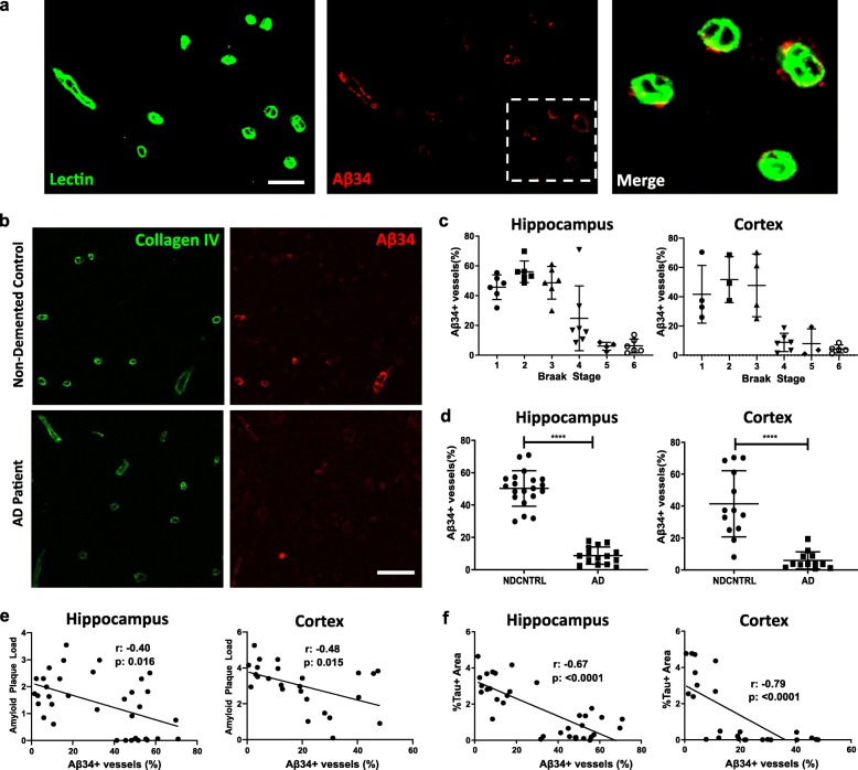
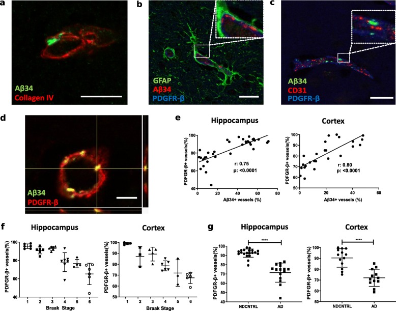
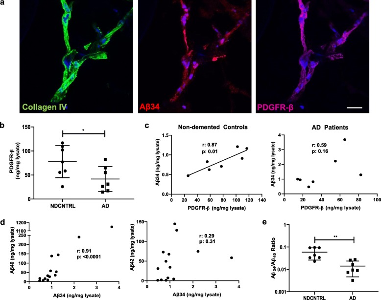
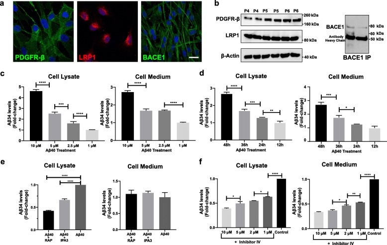
An impairment of amyloid β-peptide (Aβ) clearance is suggested to play a key role in the pathogenesis of sporadic Alzheimer's disease (AD). Amyloid degradation is mediated by various mechanisms including fragmentation by enzymes like neprilysin, matrix metalloproteinases (MMPs) and a recently identified amyloidolytic activity of β-site amyloid precursor protein cleaving enzyme 1 (BACE1). BACE1 cleavage of Aβ40 and Aβ42 results in the formation of a common Aβ34 intermediate which was found elevated in cerebrospinal fluid levels of patients at the earliest disease stages. To further investigate the role of Aβ34 as a marker for amyloid clearance in AD, we performed a systematic and comprehensive analysis of Aβ34 immunoreactivity in hippocampal and cortical post-mortem brain tissue from AD patients and non-demented elderly individuals. In early Braak stages, Aβ34 was predominantly detectable in a subset of brain capillaries associated with pericytes, while in later disease stages, in clinically diagnosed AD, this pericyte-associated Aβ34 immunoreactivity was largely lost. Aβ34 was also detected in isolated human cortical microvessels associated with brain pericytes and its levels correlated with Aβ40, but not with Aβ42 levels. Moreover, a significantly decreased Aβ34/Aβ40 ratio was observed in microvessels from AD patients in comparison to non-demented controls suggesting a reduced proteolytic degradation of Aβ40 to Aβ34 in AD. In line with the hypothesis that pericytes at the neurovascular unit are major producers of Aβ34, biochemical studies in cultured human primary pericytes revealed a time and dose dependent increase of Aβ34 levels upon treatment with recombinant Aβ40 peptides while Aβ34 production was impaired when Aβ40 uptake was reduced or BACE1 activity was inhibited. Collectively, our findings indicate that Aβ34 is generated by a novel BACE1-mediated Aβ clearance pathway in pericytes of brain capillaries. As amyloid clearance is significantly reduced in AD, impairment of this pathway might be a major driver of the pathogenesis in sporadic AD.

Keywords: Alzheimer’s disease; Amyloid clearance; Aβ34; Pericyte.


Conflict of interest statement

The authors declare that they have no competing interests.

Figures

Fig. 1
Fig. 1
Capillary associated Aβ34 immunoreactivity is reduced in Alzheimer’s disease patients and inversely correlated with both amyloid and tau. a Double immunostaining of vascular marker, Lectin (green), and Aβ34 (red) in human post-mortem brain. Merged image shows magnification of the dashed area. (Scale bar 10 μm) b Representative images of double immunostaining of basement membrane marker, Collagen IV (green) and Aβ34 (red) in a non-demented control and an AD patient for quantification of capillary Aβ34 immunoreactivity. In each visual field number of Aβ34+ vessels divided by total number of vessels (diameter < 10 μm) (calculated with Collagen IV immunostaining) and results reported as percentage of Aβ34+ vessels. (Scale bar 10 μm) c Braak-stage distribution for % of Aβ34+ vessels in hippocampus and cortex. Graphs represent individual values and mean with standard deviation (SD). d Diagnosis-based distribution of % of Aβ34+ vessels in hippocampus and cortex. Graphs represent individual values and mean with SD. **** (p < 0.001) was determined by a two-tailed unpaired student’s t test. e Correlation of %Aβ34+ vessels with amyloid plaque load in hippocampus and cortex. Amyloid plaque load was calculated by using % area covered by immunostaining of N-terminal anti-amyloid (clone W02) antibody. For hippocampus Pearson’s correlation (α = 0.05, CI 95%, two-tailed) and for cortex Spearman’s correlation were (α = 0.05, CI 95%, two-tailed) used. f Correlation of %Aβ34+ vessels with tau tangles in hippocampus and cortex. Tau+ area was calculated by using % area covered by anti-tau (clone AT8) antibody. Correlations were calculated using Spearman’s correlation (α = 0.05, CI 95%, two-tailed)
Fig. 2
Fig. 2
Aβ34 is associated with PDGFR-β + pericytes in small capillaries and Aβ34 immunoreactivity is strongly correlated with pericyte coverage a Merged image of Aβ34 (green) and basement membrane marker, Collagen IV (red) immunostaining. (Scale bar 10 μm) b Merged image of astrocyte marker, GFAP (green), Aβ34 (red) and pericyte marker, PDGFR-β (blue) immunostaining (Scale bar 20 μm). Insert shows magnification of the dashed area. c Merged image of Aβ34 (green), endothelial cell marker, CD31 (red) and pericyte marker, PDGFR-β (blue) immunostaining (Scale bar 20 μm). Insert shows magnification of the dashed area. d Maximum projection of confocal merged image of Aβ34 (green) and pericyte marker PDGFR-β (red). (Scale bar 5 μm). e Correlation of % Aβ34+ vessels with % PDGFR-β + vessels in hippocampus and cortex. % of PDGFR-β + vessels represents the number of PDGFR-β + vessels divided by total number of vessels assessed by Collagen IV immunostaining for each visual field. For hippocampus Pearson’s correlation (α = 0.05, CI 95%, two-tailed), for cortex Spearman’s correlation were (α = 0.05, CI 95%, two-tailed) used. f Braak stage distribution of %PDGFR-β + vessels (pericyte coverage) in hippocampus and cortex. Graphs represent mean with standard deviation (SD). g Diagnosis-based distribution of %PDGFR-β + vessels in hippocampus and cortex. Graphs represent individual values and mean with SD. **** (p < 0.001) was determined by a two-tailed unpaired Mann-Whitney test for hippocampus and student’s t-test for cortex
Fig. 3
Fig. 3
Strong association between pericytes and Aβ34 in microvessels isolated from frozen human cortex. a Immunostaining of basement membrane marker, Collagen IV (green), Aβ34 (red), pericyte marker, PDGFR-β (blue) and DAPI (blue) in a microvessel isolated from human cortex. (Scale bar 20 μm). b Distribution of PDGFR-β levels (normalized to total protein content) in vessel enriched brain lysates for non-demented controls and AD patients. Graph represent individual values and mean with SD. * (p < 0.05) was determined by a two-tailed unpaired student’s t test. c Correlation of Aβ34 and PDGFR-β levels (normalized to total protein content) in vessel enriched brain lysates in non-demented controls and AD patients. Correlations were calculated using Pearson’s correlation (α = 0.05, CI 95%, two-tailed). d Correlation of Aβ34 and Aβ40 or Aβ42 levels (normalized to total protein content) in vessel enriched brain lysates. For Aβ34 and Aβ40 correlation, Spearman’s correlation (α = 0.05, CI 95%, two-tailed) was used. For Aβ34 and Aβ42 correlation, Pearson’s correlation (α = 0.05, CI 95%, two-tailed) was used. e Distribution of Aβ34/Aβ40 ratio in vessel enriched brain lysates for non-demented controls and AD patients. Graph represent individual values and mean with SD. ** (p < 0.01) was determined by a two-tailed unpaired Mann-Whitney test
Fig. 4
Fig. 4
Human primary pericytes take up extracellular Aβ40 via LRP1-mediated endocytosis and generate Aβ34 via BACE1-mediated enzymatic degradation. a Immunostaining of human primary pericytes with PDGFR-β, LRP1, BACE1 and DAPI (blue). (Scale bar 10 μm) b Levels of PDGFR-β and LRP1 in pericyte lysates from different passages (passage 4 (P4), passage 5 (P5) and passage 6 (P6)) assessed by western blot. Levels of BACE1 in pericyte lysate were also assessed by western blot following immunoprecipitation (IP). c Aβ34 levels in cell lysate and medium following 3 days incubation with varying concentrations of human recombinant Aβ40 peptide. Aβ34 levels were normalized and shown as fold-change compared to lowest concentration of Aβ40 treatment (1 μM). d Aβ34 levels in cell lysate and medium following incubation with 5 μM human recombinant Aβ40 peptide for varying durations. Aβ34 levels were normalized and shown as fold-change compared to shortest duration of Aβ40 treatment (12 h). e Aβ34 levels in cell lysate and medium following 2 h incubation with 10 μM IPA3 or 500 nM RAP prior to 12 h incubation with 2 μM Aβ40. Aβ34 levels were normalized and shown as fold-change compared to only Aβ40 treated cells. f Aβ34 levels in cell lysate and medium following 12 h incubation with varying concentrations of BACE1 inhibitor (Inhibitor IV) prior to 12 h incubation with 2 μM Aβ40. Aβ34 levels were normalized and shown as fold-change compared to only Aβ40 treated cells. The graphs (c-f) represent the mean ± SD. **** (p < 0.0001), *** (p < 0.001), ** (p < 0.01) and * (p < 0.05) were determined by 1-way ANOVA followed by Tukey’s multiple comparison test

Similar articles

Cited by

References

    1. Armulik A, Genové G, Betsholtz C. Pericytes: developmental, physiological, and pathological perspectives, problems, and promises. Dev Cell. 2011;21:193–215. doi: 10.1016/j.devcel.2011.07.001. - DOI - PubMed
    1. Armulik A, Genové G, Mäe M, Nisancioglu MH, Wallgard E, Niaudet C, He L, Norlin J, Lindblom P, Strittmatter K, Johansson BR, Betsholtz C. Pericytes regulate the blood-brain barrier. Nature. 2010;468:557–561. doi: 10.1038/nature09522. - DOI - PubMed
    1. Bancher C, Braak H, Fischer P, Jellinger KA. Neuropathological staging of Alzheimer lesions and intellectual status in Alzheimer’s and Parkinson’s disease patients. Neurosci Lett. 1993;162:179–182. doi: 10.1016/0304-3940(93)90590-H. - DOI - PubMed
    1. Bell RD, Winkler EA, Sagare AP, Singh I, LaRue B, Deane R, Zlokovic BV. Pericytes control key neurovascular functions and neuronal phenotype in the adult brain and during brain aging. Neuron. 2010;68:409–427. doi: 10.1016/j.neuron.2010.09.043. - DOI - PMC - PubMed
    1. Bigl M, Apelt J, Luschekina EA, Lange-Dohna C, Rossner S, Schliebs R. Expression of beta-secretase mRNA in transgenic Tg2576 mouse brain with Alzheimer plaque pathology. Neurosci Lett. 2000;292:107–110. doi: 10.1016/s0304-3940(00)01452-x. - DOI - PubMed

Publication types

MeSH terms