. 2019 Dec 3;12(1):102.
doi: 10.1186/s13041-019-0523-7.

Silencing of microRNA-146a alleviates the neural damage in temporal lobe epilepsy by down-regulating Notch-1

Affiliations

Silencing of microRNA-146a alleviates the neural damage in temporal lobe epilepsy by down-regulating Notch-1

Hui Huang et al. Mol Brain. .

Abstract

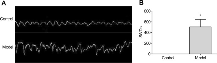
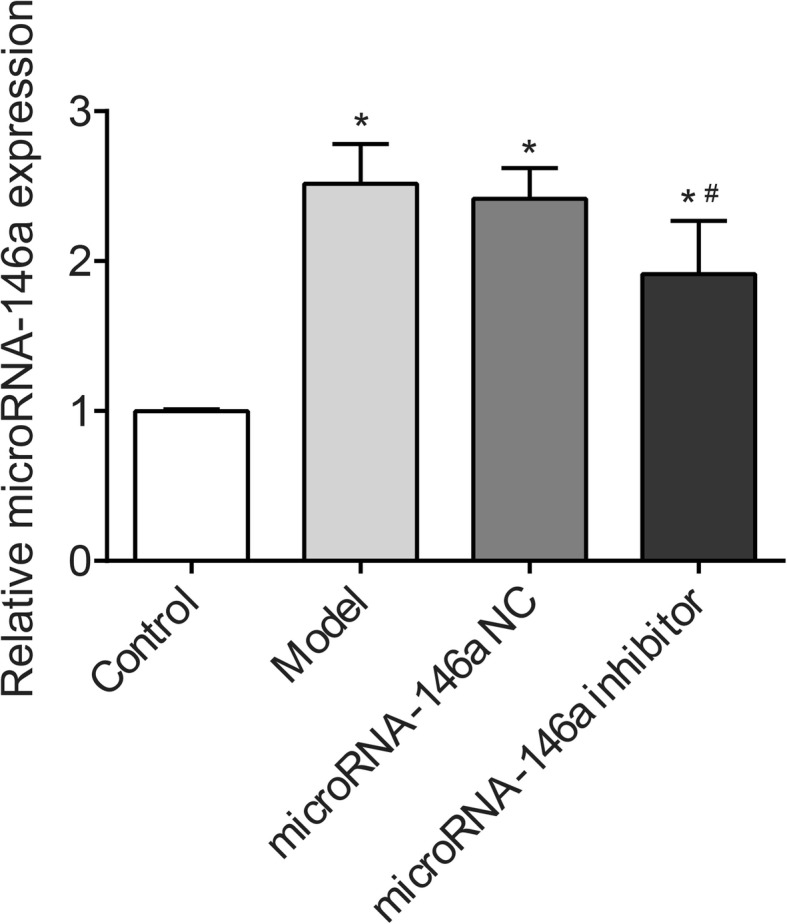
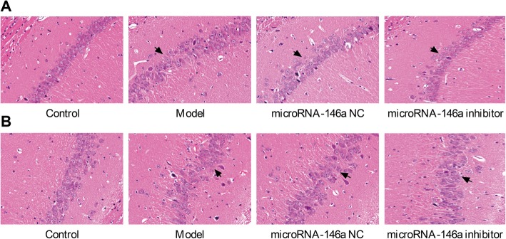
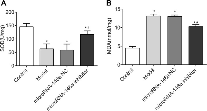
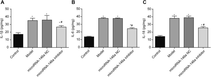
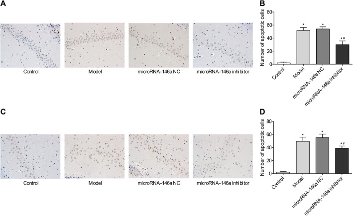
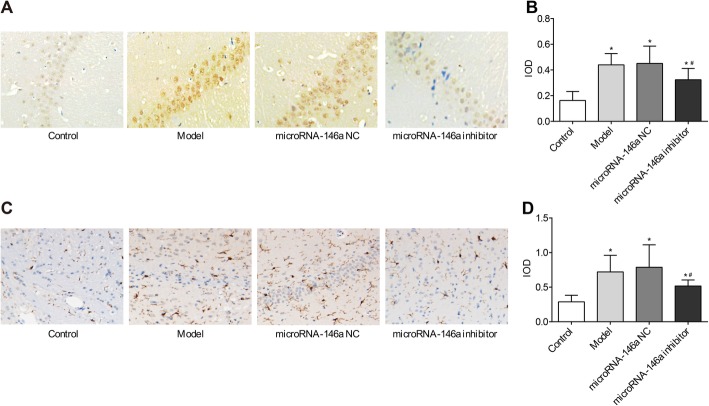
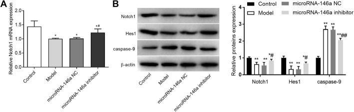
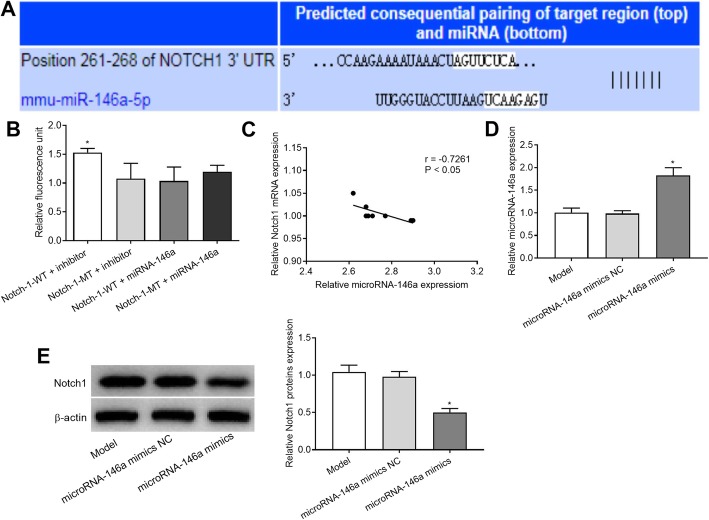
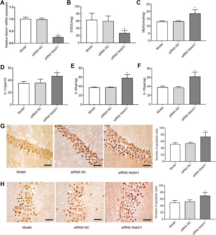
This study aimed to evaluate the specific regulatory roles of microRNA-146a (miRNA-146a) in temporal lobe epilepsy (TLE) and explore the related regulatory mechanisms. A rat model of TLE was established by intraperitoneal injection of lithium chloride-pilocarpine. These model rats were injected intracerebroventricularly with an miRNA-146a inhibitor and Notch-1 siRNA. Then, neuronal damage and cell apoptosis in the cornu ammonis (CA) 1 and 3 regions of the hippocampus were assessed. SOD and MDA levels in the hippocampus were detected by chromatometry, and IL-1β, IL-6, and IL-18 levels were detected by ELISA. Then, we evaluated the expression levels of caspase-9, GFAP, Notch-1, and Hes-1 in the hippocampus. The interaction between Notch-1 and miRNA-146a was assessed by a dual luciferase reporter gene assay. A rat model of TLE was successfully established, which exhibited significantly increased miRNA-146a expression in the hippocampus. Silencing of miRNA-146a significantly alleviated the neuronal damage and cell apoptosis in the CA1 and CA3 regions of the hippocampus in TLE rats and decreased MDA, IL-1β, IL-6, and IL-18 levels and increased SOD levels in the hippocampus of TLE rats. In addition, silencing of miRNA-146a significantly decreased the expression levels of caspase-9, GFAP, Notch-1, and Hes-1 in the hippocampus of TLE rats. Notch-1 was identified as a target of miRNA-146a and silencing of Notch-1 aggravated the neuronal damage in the CA1 and CA3 regions. Silencing of miRNA-146a alleviated the neuronal damage in the hippocampus of TLE rats by down-regulating Notch-1.

Keywords: Caspase-9; Neuronal damage; Notch-1; Temporal lobe epilepsy; microRNA-146a.


Conflict of interest statement

The authors declare that they have no competing interests.

Figures

Fig. 1
Fig. 1
The electroencephalograms (EEGs) of rats were measured after 24 h post-epilepsy occurrence. Model, temporal lobe epilepsy (TLE) rats; Control, normal rats. a EEGs; b) the number of spike-and-wave discharges (SWDs). *, P < 0.05 vs. Control group
Fig. 2
Fig. 2
The expression of microRNA-146a (miRNA-146a) in hippocampus of rats detected by quantitative real-time PCR (qRT-PCR) (N = 8) at 3 days after the injection. Model, temporal lobe epilepsy (TLE) rats; Control, normal rats. *, P < 0.05 vs. Control group; #, P < 0.05 vs. Model group
Fig. 3
Fig. 3
Pathological morphology of cornu ammonis (CA)1 and CA3 regions in hippocampus of rats were detected by hematoxylin-eosin (HE) staining (× 400) (N = 8) at 3 days after the injection. Model, temporal lobe epilepsy (TLE) rats; Control, normal rats; microRNA-146a (miRNA-146a) siRNA, TLE rats transfected with miRNA-146a siRNA; miRNA-146a negative control (miRNA-146a NC), TLE rats transfected with miRNA-146a NC. a CA1 region; b) CA3 region. Arrows represented the accumulation area of neurons
Fig. 4
Fig. 4
The levels of SOD and MDA in hippocampus of rats were detected by chromatometry (N = 8) at 3 days after the injection. Model, temporal lobe epilepsy (TLE) rats; Control, normal rats; microRNA-146a (miRNA-146a) siRNA, TLE rats transfected with miRNA-146a siRNA; miRNA-146a negative control (miRNA-146a NC), TLE rats transfected with miRNA-146a NC. a SOD level; b) MDA level. *, P < 0.05 vs. Control group; #, P < 0.05 vs. Model group
Fig. 5
Fig. 5
The levels of IL-1β, IL-6, and IL-18 in hippocampus of rats were detected by enzyme linked immunosorbent assay (ELISA) (N = 8) at 3 days after the injection. Model, temporal lobe epilepsy (TLE) rats; Control, normal rats; microRNA-146a (miRNA-146a) siRNA, TLE rats transfected with miRNA-146a siRNA; miRNA-146a negative control (miRNA-146a NC), TLE rats transfected with miRNA-146a NC. a IL-1β; b) IL-6; c) IL-18. *, P < 0.05 vs. Control group; #, P < 0.05 vs. Model group
Fig. 6
Fig. 6
Cell apoptosis in CA1 and CA3 regions of hippocampus in rats detected by TUNEL (N = 5). Model, temporal lobe epilepsy (TLE) rats; Control, normal rats; microRNA-146a (miRNA-146a), TLE rats transfected with miRNA-146a siRNA; miRNA-146a negative control (miRNA-146a NC), TLE rats transfected with miRNA-146a NC. a microscopic observation of apoptotic cells in CA1 region; b) the number of apoptotic cells in stratum pyramidale of CA1 region; c) microscopic observation of apoptotic cells in CA3 region; d) the number of apoptotic cells in stratum pyramidale of CA3 region. *, P < 0.05 vs. Control group; #, P < 0.05 vs. Model group
Fig. 7
Fig. 7
The expression of caspase-9 and GFAP in hippocampus was detected by immunohistochemistry (ICH) (× 400) (N = 8) at 3 days after the injection. Model, temporal lobe epilepsy (TLE) rats; Control, normal rats; microRNA-146a (miRNA-146a), TLE rats transfected with miRNA-146a siRNA; miRNA-146a negative control (miRNA-146a NC), TLE rats transfected with miRNA-146a NC. a microscopic observation of caspase-9-positive cells; b) the expression level of caspase-9; c) microscopic observation of GFAP-positive cells; d) the expression level of GFAP. *, P < 0.05 vs. Control group; #, P < 0.05 vs. Model group
Fig. 8
Fig. 8
The expression of Notch-1, Hes-1 and caspase-9 in hippocampus detected by quantitative real-time PCR (qRT-PCR) and/or Western blot (N = 8) was performed at 3 days after the injection. Model, temporal lobe epilepsy (TLE) rats; Control, normal rats; microRNA-146a (miRNA-146a), TLE rats transfected with miRNA-146a siRNA; miRNA-146a negative control (miRNA-146a NC), TLE rats transfected with miRNA-146a NC. a the expression of Notch-1 detected by qRT-PCR (mRNA level); b) the expression of Notch-1, Hes-1 and caspase-9 detected by Western blot (protein level). *, P < 0.05 vs. Control group; **, P < 0.01 vs. Control group; #, P < 0.05 vs. Model group
Fig. 9
Fig. 9
The interaction between Notch-1 and microRNA-146a (miRNA-146a). a a binding site of miRNA-146a at 3′-UTR of Notch-1 predicted by Target Scan; b) the relative fluorescence unit of co-transfected cells detected by dual luciferase reporter gene (DLR) assay. *, P < 0.05 vs. Notch-1-WT + miRNA-146a inhibitor. c Spearman’s rank correlation analysis was used to detect the correlation between Notch1 and miRNA-146a expression. d The expression of miRNA-146a transfected with miRNA-146a mimics or miRNA-146a mimics NC in the hippocampus of rats was detected by qRT-PCR. e Western blot analysis was used to detect the expression of Notch1 protein after transfected with miRNA-146a mimics or miRNA-146a mimics NC in the hippocampus of rats. *, P < 0.05 vs. miRNA-146a mimics NC
Fig. 10
Fig. 10
Silence Notch1 aggravated the severity of epileptic brain damage. a Rats (N = 8 in each group) were injected intracerebroventricularly with siRNA negative control (siRNA NC) or siRNA targeting Notch1 (siRNA Notch1) for three days and relative Notch1 expression was assessed by qRT-PCR to detect the siRNA efficiency against Notch1. After transfection of 3 days, the levels of SOD (b), MDA (c), IL-1β (d), IL-6 (e) and IL-18 (f) were detected. *P < 0.05 vs. siNC group. Photomicrographs of apoptotic cells in CA1 region (g) and (h) CA3 region (arrows indicate the positive cells, bar = 100 μm, magnification × 400)

References

    1. Raoof R, Jimenez-Mateos EM, Bauer S, Tackenberg B, Rosenow F, Lang J, et al. Cerebrospinal fluid microRNAs are potential biomarkers of temporal lobe epilepsy and status epilepticus. Sci Rep. 2017:7. - PMC - PubMed
    1. Spencer SS. Neural networks in human epilepsy: evidence of and implications for treatment. Epilepsia. 2010;43:219–227. doi: 10.1046/j.1528-1157.2002.26901.x. - DOI - PubMed
    1. Thom M, Eriksson S, Martinian L, Caboclo LO, Mcevoy AW, Duncan JS, et al. Temporal lobe sclerosis associated with hippocampal sclerosis in temporal lobe epilepsy: Neuropathological features. J Neuropathol Exp Neurol. 2009;68:928–938. doi: 10.1097/NEN.0b013e3181b05d67. - DOI - PMC - PubMed
    1. Coyle A, Riley J, Wu C, Sharan A. From resection to ablation: a review of resective surgical options for temporal lobe epilepsy and rationale for an ablation-based approach. Neurol India. 2017;65:S71. - PubMed
    1. Bonilha L, Keller SS. Quantitative MRI in refractory temporal lobe epilepsy: relationship with surgical outcomes. Quant Imaging Med Surg. 2015;5:204. - PMC - PubMed

MeSH terms

LinkOut - more resources