. 2019 Dec 3;7(1):192.
doi: 10.1186/s40478-019-0823-2.

A post-translational modification signature defines changes in soluble tau correlating with oligomerization in early stage Alzheimer's disease brain

Affiliations

A post-translational modification signature defines changes in soluble tau correlating with oligomerization in early stage Alzheimer's disease brain

Ebru Ercan-Herbst et al. Acta Neuropathol Commun. .

Abstract

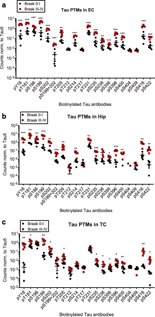
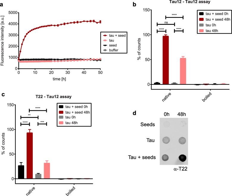
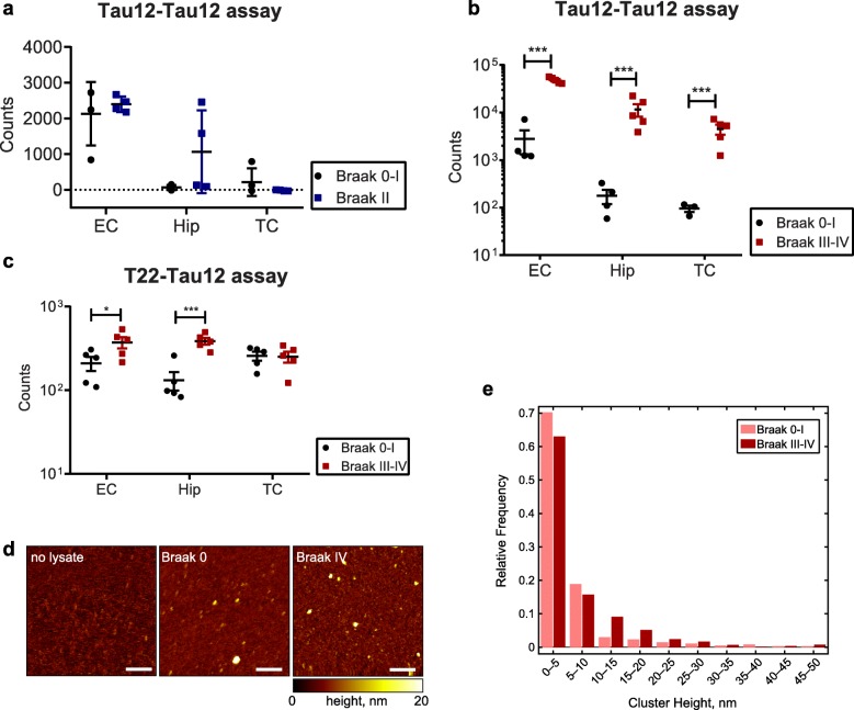
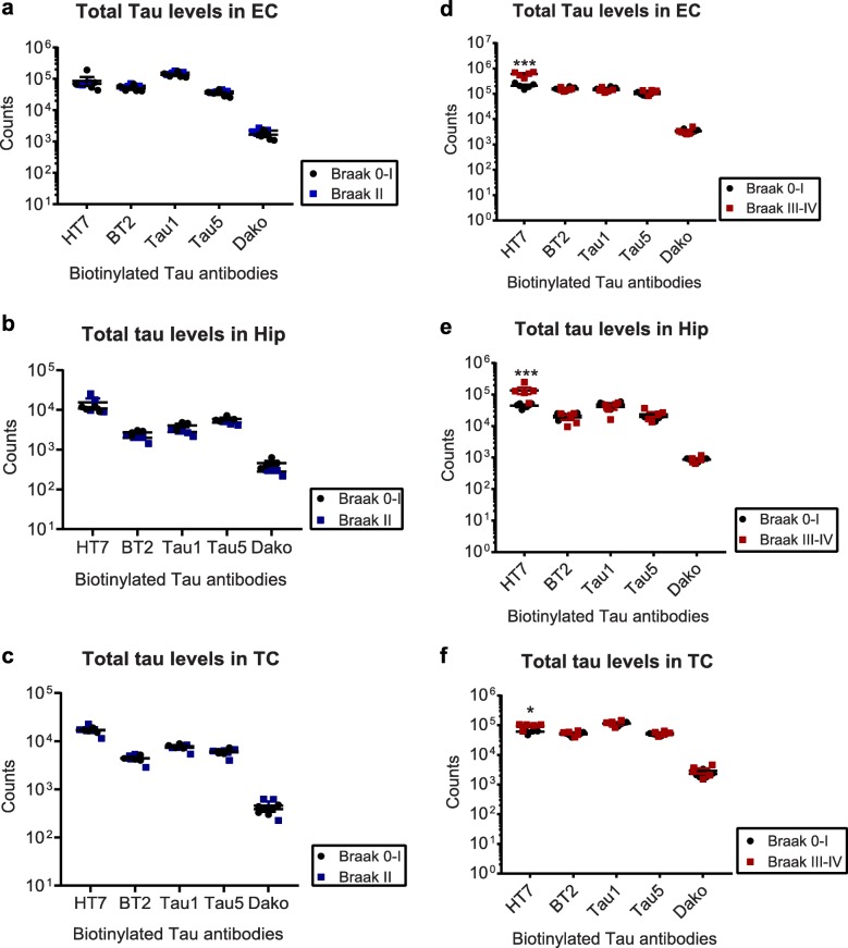
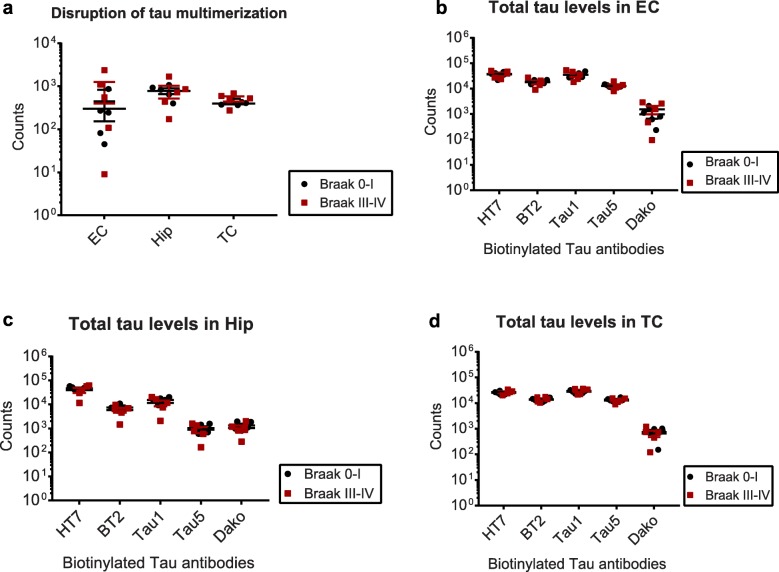
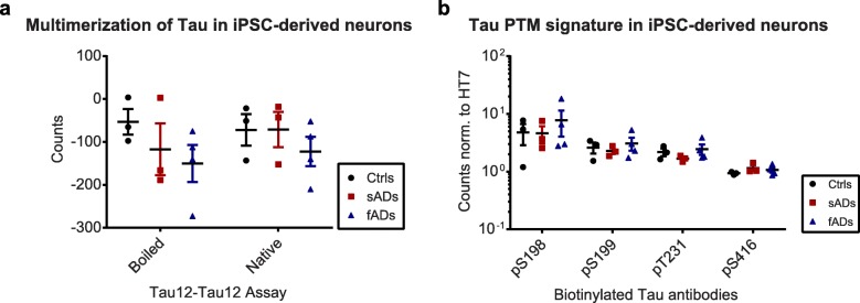
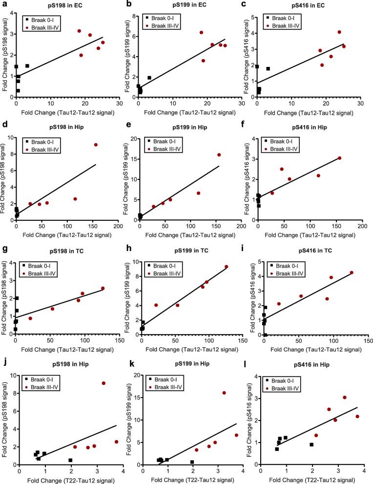
Tau is a microtubule-binding protein that can receive various post-translational modifications (PTMs) including phosphorylation, methylation, acetylation, glycosylation, nitration, sumoylation and truncation. Hyperphosphorylation of tau is linked to its aggregation and the formation of neurofibrillary tangles (NFTs), which are a hallmark of Alzheimer's disease (AD). While more than 70 phosphorylation sites have been detected previously on NFT tau, studies of oligomeric and detergent-soluble tau in human brains during the early stages of AD are lacking. Here we apply a comprehensive electrochemiluminescence ELISA assay to analyze twenty-five different PTM sites as well as tau oligomerization in control and sporadic AD brain. The samples were classified as Braak stages 0-I, II or III-IV, corresponding to the progression of microscopically detectable tau pathology throughout different brain regions. We found that soluble tau multimers are strongly increased at Braak stages III-IV in all brain regions under investigation, including the temporal cortex, which does not contain NFTs or misfolded oligomers at this stage of pathology. We additionally identified five phosphorylation sites that are specifically and consistently increased across the entorhinal cortex, hippocampus and temporal cortex in the same donors. Three of these sites correlate with tau multimerization in all three brain regions, but do not overlap with the epitopes of phospho-sensitive antibodies commonly used for the immunohistochemical detection of NFTs. Our results thus suggest that soluble multimers are characterized by a small set of specific phosphorylation events that differ from those dominating in mature NFTs. These findings shed light on early PTM changes of tau during AD pathogenesis in human brains.

Keywords: Alzheimer’s disease; Posttranslational modifications; Tau; Tau oligomerization.


Conflict of interest statement

The authors declare that they have no competing interests.

Figures

Fig. 1
Fig. 1
Tau phosphorylation does not change in Braak II samples compared to Braak 0–I controls. Normalized phospho-tau signals from Braak II and Braak 0–I a) entorhinal cortices (EC), b) Hippocampi (Hip) and c) Temporal cortices (TC). Biotinylated antibodies were used as capture, sulfo-tagged Tau12 was used for detection. None of the observed changes were significant (p > 0.05, t-tests)
Fig. 2
Fig. 2
Compared to Braak 0–I samples, many but not all, tau phosphorylation events are increased in native Braak III–IV samples. Normalized phospho-tau signals obtained from ELISA measurements of samples from a) entorhinal cortices (EC), b) hippocampi (Hip) and c) temporal cortices (TC). Student’s t-tests: *, p < 0.05, **, p < 0.01, ***, p < 0.001
Fig. 3
Fig. 3
Oligomerization of tau can be monitored with Tau12-Tau12 or T22-Tau12 ELISA. a) Fluorescence intensities of ThT assays showing aggregation of recombinant full-length tau over time. Seeds alone (tau aa256–368), buffer alone and full-length tau alone were used as controls. The signal for tau with seeds increases exponentially until app. 8 h of incubation (n = 3). Analysis of aggregates by b) Tau12-Tau12 ELISA assay and c) T22-Tau12 ELISA assay. Both methods yield a higher signal for tau with seeds after 48 h of incubation, which is abolished after boiling in SDS-containing buffer (n = 3). d) Dot blot analysis of native samples with T22 antibody: seeds alone, tau alone and tau with seeds at 0 h and 48 h. Two-way Anova for b and c: **, p < 0.01, ***, p < 0.001, ****, p < 0.0001
Fig. 4
Fig. 4
ELISA assays and atomic force microscopy (AFM) reveal more abundant tau oligomers in Braak III–IV ECs. Comparison of ELISA counts from Braak 0–I controls with a) Braak II entorhinal cortices (EC), hippocampi (Hip), and temporal cortices (TC) and b, c) Braak III–IV EC, Hip and TC, using Tau12-Tau12 (a, b) or T22-Tau12 (c) assays. d) Representative AFM images of eluates after Tau12 immunoprecipitation; left: eluate without brain lysate (negative control), middle: eluate of Braak 0 EC, right: eluate of Braak IV EC. Scale bars represent 200 nm. e) Relative frequencies of cluster heights detected from AFM scans of two Braak 0 and two Braak IV EC tissue samples shows increase of clusters > 10 nm in Braak IV samples. Number of clusters detected: Braak 0–I: 1343, Braak III–IV: 1053. Student’s t-tests: *, p < 0.05, ***, p < 0.001
Fig. 5
Fig. 5
Total tau levels at different Braak stages in different brain regions. Total tau levels in a, d) Entorhinal cortices (EC), b, e) Hippocampi (Hip), and c, f) Temporal Cortices (TC) from Braak II (a–c) and Braak III–IV (d–f) samples, along with their age-matched Braak 0–I controls. Student’s t-tests: *, p < 0.05, ***, p < 0.001
Fig. 6
Fig. 6
Tau multimers can be disrupted by boiling in SDS-containing buffer. a) Comparison of tau multimer levels in entorhinal cortices, hippocampi, and temporal cortices between Braak 0–I and Braak III–IV after boiling with SDS-containing buffer. Comparison of total tau levels in b) Entorhinal cortices (EC) c) Hippocampi (Hip) and d) Temporal cortices (TC) between Braak 0–I and Braak III–IV
Fig. 7
Fig. 7
Tau PTMs in denatured Braak III–IV samples. Normalized PTM signals from a) Entorhinal cortices (EC), c) hippocampi (Hip), and e) temporal cortices (TC) of Braak stages 0–I and III–IV. b, d, f) Corresponding fold changes (log2) versus significance (−log10(p-value)) of the changes. Phosphorylation at the sites above the red line, which corresponds to p-value = 0.05, is significantly higher in Braak III–IV samples
Fig. 8
Fig. 8
iPSC-derived neurons do not recapitulate the tau PTM signature. a) Analysis of multimers by Tau12-Tau12 electrochemiluminescence assay with and without boiling of lysates from controls, familial AD (fAD) and sporadic AD (sAD) neurons with SDS. b) Normalized PTM signals (pS198, pS199, pT231 and pS416). None of the observed changes were significant (p > 0.05, t-tests)
Fig. 9
Fig. 9
Correlation of tau oligomerization with pS198, pS199, and pS416 fold changes in all brain regions. Spearman correlation of the fold changes in Tau12-Tau12 signal with the fold changes (black squares: Braak 0–I / average (Braak 0–I); red circles: Braak III–IV / average (Braak 0–I)) of a) pS198, b) pS199 and c) pS416 in entorhinal cortex (EC), d) pS198, e) pS199 and f) pS416 in hippocampus (Hip), g) pS198, h) pS199 and i) pS416 in temporal cortex (TC) and Spearman correlation of the fold changes in T22-Tau12 signal with the fold changes (black squares: Braak 0–I / average (Braak 0–I), red circles: Braak III–IV / average (Braak 0–I)) of j) pS198, k) pS199 and l) pS416 in hippocampus (Hip). Results of the statistical analysis are summarized in Table 4

References

    1. Alzheimer A (1907) Über eine eigenartige Erkrankung der Hirnrinde. Allgemeine Zeitschrift fur Psychiatrie und Psychisch-gerichtliche Medizin 64: 146–148
    1. Ince PG, McArthur FK, Bjertness E, Torvik A, Candy JM, Edwardson JA (1995) Neuropathological diagnoses in elderly patients in Oslo: Alzheimer's disease, Lewy body disease, vascular lesions. Dementia 6: 162–168 - PubMed
    1. Bowler JV, Munoz DG, Merskey H, Hachinski V (1998) Fallacies in the pathological confirmation of the diagnosis of Alzheimer's disease. J Neurol Neurosurg Psychiatry 64: 18–24 - PMC - PubMed
    1. Dickson DW (1997) Neuropathological diagnosis of Alzheimer's disease: a perspective from longitudinal clinicopathological studies. Neurobiol Aging 18: S21–26 - PubMed
    1. Braak H, Braak E (1998) Evolution of neuronal changes in the course of Alzheimer's disease. J Neural Transm Suppl 53: 127–140 - PubMed

Publication types