. 2019 Dec 3;10(1):5511.
doi: 10.1038/s41467-019-13494-7.

Cryo-EM structure of a 40 kDa SAM-IV riboswitch RNA at 3.7 Å resolution

Affiliations

Cryo-EM structure of a 40 kDa SAM-IV riboswitch RNA at 3.7 Å resolution

Kaiming Zhang et al. Nat Commun. .

Abstract

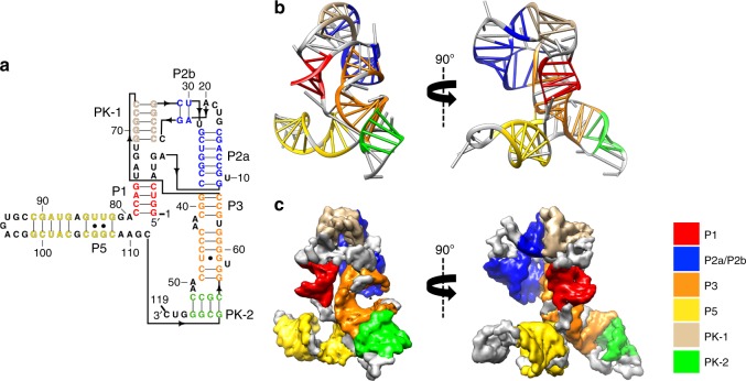
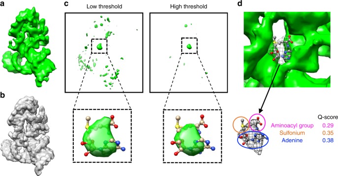
Specimens below 50 kDa have generally been considered too small to be analyzed by single-particle cryo-electron microscopy (cryo-EM). The high flexibility of pure RNAs makes it difficult to obtain high-resolution structures by cryo-EM. In bacteria, riboswitches regulate sulfur metabolism through binding to the S-adenosylmethionine (SAM) ligand and offer compelling targets for new antibiotics. SAM-I, SAM-I/IV, and SAM-IV are the three most commonly found SAM riboswitches, but the structure of SAM-IV is still unknown. Here, we report the structures of apo and SAM-bound SAM-IV riboswitches (119-nt, ~40 kDa) to 3.7 Å and 4.1 Å resolution, respectively, using cryo-EM. The structures illustrate homologies in the ligand-binding core but distinct peripheral tertiary contacts in SAM-IV compared to SAM-I and SAM-I/IV. Our results demonstrate the feasibility of resolving small RNAs with enough detail to enable detection of their ligand-binding pockets and suggest that cryo-EM could play a role in structure-assisted drug design for RNA.


Conflict of interest statement

The authors declare no competing interests.

Figures

Fig. 1
Fig. 1
Single-particle cryo-EM analysis of the apo SAM-IV riboswitch collected on Titan Krios. a Representative motion-corrected cryo-EM micrograph. Scale bar represents 100 Å. b Reference-free 2D class averages computed in Relion. c Final 3D reconstruction in two different views.
Fig. 2
Fig. 2
The secondary and tertiary structures of the apo SAM-IV riboswitch. a The secondary structure with the domains colored differently. The black arrow indicates the direction of the backbone. b-c The model and map colored by the same scheme as in a. P paired, PK pseudoknot.
Fig. 3
Fig. 3
Determination of the ligand location in the ligand-bound SAM-IV cryo-EM map. a Ligand-bound SAM-IV cryo-EM map. b Map computed from the SAM-IV RNA-only model from a. c Difference map between a and b shown at two different thresholds. The insets show the prominent density in the difference map into which are fitted the ligand model derived from SAM-I crystal structure (PDB code: 3GX5). d Segmented SAM density from a is highlighted in a wire frame representation, fitted with the ligand model derived from SAM-I crystal structure (PDB code: 3GX5). Q-scores of the ligand components between the map and model are calculated.

References

    1. Zhang K, et al. Structure of the 30 kDa HIV-1 RNA dimerization signal by a hybrid Cryo-EM, NMR, and molecular dynamics approach. Structure. 2018;26:490–498.e3. doi: 10.1016/j.str.2018.01.001. - DOI - PMC - PubMed
    1. Roth A, Breaker RR. The structural and functional diversity of metabolite-binding riboswitches. Annu. Rev. Biochem. 2009;78:305–334. doi: 10.1146/annurev.biochem.78.070507.135656. - DOI - PMC - PubMed
    1. Howe JA, et al. Atomic resolution mechanistic studies of ribocil: a highly selective unnatural ligand mimic of theE. coliFMN riboswitch. RNA Biol. 2016;13:946–954. doi: 10.1080/15476286.2016.1216304. - DOI - PMC - PubMed
    1. Connelly CM, Moon MH, Schneekloth JS., Jr The emerging role of RNA as a therapeutic target for small molecules. Cell Chem. Biol. 2016;23:1077–1090. doi: 10.1016/j.chembiol.2016.05.021. - DOI - PMC - PubMed
    1. Mulhbacher J, et al. Novel riboswitch ligand analogs as selective inhibitors of guanine-related metabolic pathways. PLoS Pathog. 2010;6:e1000865. doi: 10.1371/journal.ppat.1000865. - DOI - PMC - PubMed

Publication types