

Prenatal Glucocorticoid Exposure Results in Changes in Gene Transcription and DNA Methylation in the Female Juvenile Guinea Pig Hippocampus Across Three Generations
- PMID: 31796763
- PMCID: PMC6890750
- DOI: 10.1038/s41598-019-54456-9
Prenatal Glucocorticoid Exposure Results in Changes in Gene Transcription and DNA Methylation in the Female Juvenile Guinea Pig Hippocampus Across Three Generations
Abstract
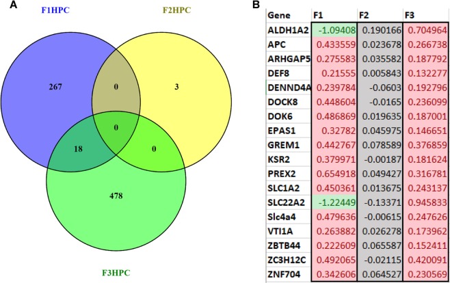
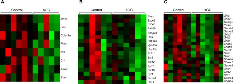
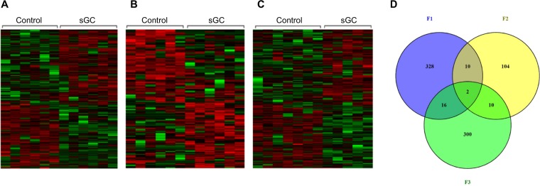
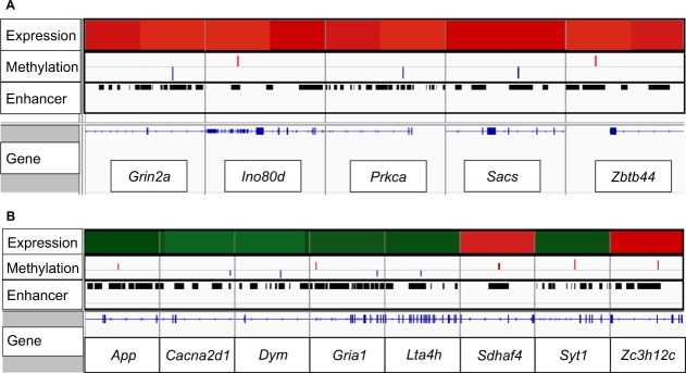
Synthetic glucocorticoids (sGC) are administered to women at risk for pre-term delivery, to mature the fetal lung and decrease neonatal morbidity. sGC also profoundly affect the fetal brain. The hippocampus expresses high levels of glucocorticoid (GR) and mineralocorticoid receptor (MR), and its development is affected by elevated fetal glucocorticoid levels. Antenatal sGC results in neuroendocrine and behavioral changes that persist in three generations of female guinea pig offspring of the paternal lineage. We hypothesized that antenatal sGC results in transgenerational changes in gene expression that correlate with changes in DNA methylation. We used RNASeq and capture probe bisulfite sequencing to investigate the transcriptomic and epigenomic effects of antenatal sGC exposure in the hippocampus of three generations of juvenile female offspring from the paternal lineage. Antenatal sGC exposure (F0 pregnancy) resulted in generation-specific changes in hippocampal gene transcription and DNA methylation. Significant changes in individual CpG methylation occurred in RNApol II binding regions of small non-coding RNA (snRNA) genes, which implicates alternative splicing as a mechanism involved in transgenerational transmission of the effects of antenatal sGC. This study provides novel perspectives on the mechanisms involved in transgenerational transmission and highlights the importance of human studies to determine the longer-term effects of antenatal sGC on hippocampal-related function.
Conflict of interest statement
The authors declare no competing interests.
Figures
Similar articles
-
Adult glucocorticoid exposure leads to transcriptional and DNA methylation changes in nuclear steroid receptors in the hippocampus and kidney of mouse male offspring.Biol Reprod. 2014 Feb 27;90(2):43. doi: 10.1095/biolreprod.113.115899. Print 2014 Feb. Biol Reprod. 2014. PMID: 24451982
-
DNA methylome signatures of prenatal exposure to synthetic glucocorticoids in hippocampus and peripheral whole blood of female guinea pigs in early life.Transl Psychiatry. 2021 Jan 18;11(1):63. doi: 10.1038/s41398-020-01186-6. Transl Psychiatry. 2021. PMID: 33462183 Free PMC article.
-
Effects of antenatal synthetic glucocorticoid on glucocorticoid receptor binding, DNA methylation, and genome-wide mRNA levels in the fetal male hippocampus.Endocrinology. 2013 Nov;154(11):4170-81. doi: 10.1210/en.2013-1484. Epub 2013 Sep 12. Endocrinology. 2013. PMID: 24029241
-
Gestational arsenic exposure and paternal intergenerational epigenetic inheritance.Toxicol Appl Pharmacol. 2020 Dec 15;409:115319. doi: 10.1016/j.taap.2020.115319. Epub 2020 Nov 6. Toxicol Appl Pharmacol. 2020. PMID: 33160984 Review.
-
Glucocorticoids and fetal programming part 2: Mechanisms.Nat Rev Endocrinol. 2014 Jul;10(7):403-11. doi: 10.1038/nrendo.2014.74. Epub 2014 May 27. Nat Rev Endocrinol. 2014. PMID: 24863383 Review.
Cited by
-
Metabolic Consequences of Glucocorticoid Exposure before Birth.Nutrients. 2022 May 30;14(11):2304. doi: 10.3390/nu14112304. Nutrients. 2022. PMID: 35684104 Free PMC article. Review.
-
Placental CRH as a Signal of Pregnancy Adversity and Impact on Fetal Neurodevelopment.Front Endocrinol (Lausanne). 2021 Aug 2;12:714214. doi: 10.3389/fendo.2021.714214. eCollection 2021. Front Endocrinol (Lausanne). 2021. PMID: 34408727 Free PMC article. Review.
-
Cardinal role of the environment in stress induced changes across life stages and generations.Neurosci Biobehav Rev. 2021 May;124:137-150. doi: 10.1016/j.neubiorev.2021.01.012. Epub 2021 Feb 4. Neurosci Biobehav Rev. 2021. PMID: 33549740 Free PMC article. Review.
-
Epigenetic effects of endogenous and exogenous glucocorticosteroids during pregnancy on the offspring: a systematic-narrative review.Hormones (Athens). 2025 Jun 21. doi: 10.1007/s42000-025-00671-1. Online ahead of print. Hormones (Athens). 2025. PMID: 40542332 Review.
-
Parental mutations influence wild-type offspring via transcriptional adaptation.Sci Adv. 2022 Nov 25;8(47):eabj2029. doi: 10.1126/sciadv.abj2029. Epub 2022 Nov 25. Sci Adv. 2022. PMID: 36427314 Free PMC article.
References
-
- Bielas H, Arck P, Bruenahl CA, Walitza S, Grunblatt E. Prenatal stress increases the striatal and hippocampal expression of correlating c-FOS and serotonin transporters in murine offspring. International Journal of Developmental Neuroscience. 2014;38:30–35. doi: 10.1016/j.ijdevneu.2014.07.006. - DOI - PubMed
-
- Crudo, A. et al. Glucocorticoid programming of the fetal male hippocampal epigenome. Endocrinology154 (2013). - PubMed
Publication types
MeSH terms
Substances
Grants and funding
LinkOut - more resources
Full Text Sources
Medical
Molecular Biology Databases
