

Honey can inhibit and eliminate biofilms produced by Pseudomonas aeruginosa
- PMID: 31796774
- PMCID: PMC6890799
- DOI: 10.1038/s41598-019-54576-2
Honey can inhibit and eliminate biofilms produced by Pseudomonas aeruginosa
Abstract
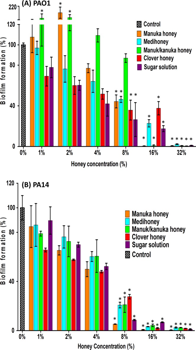
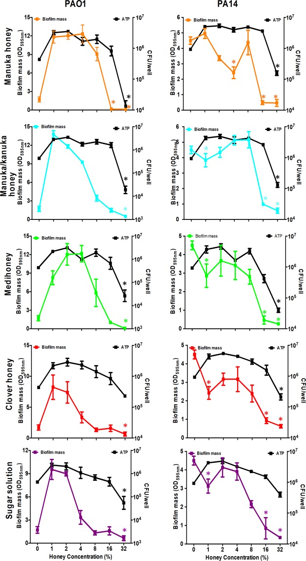
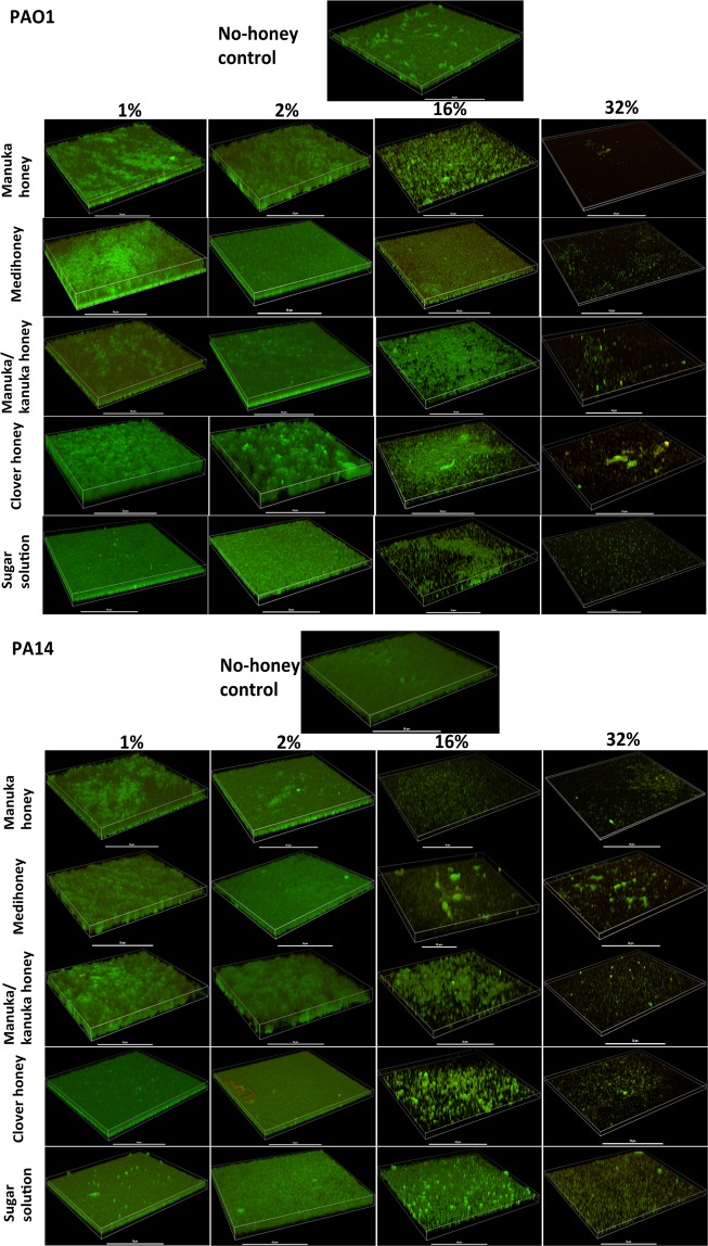
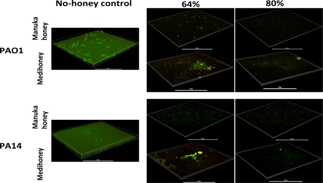
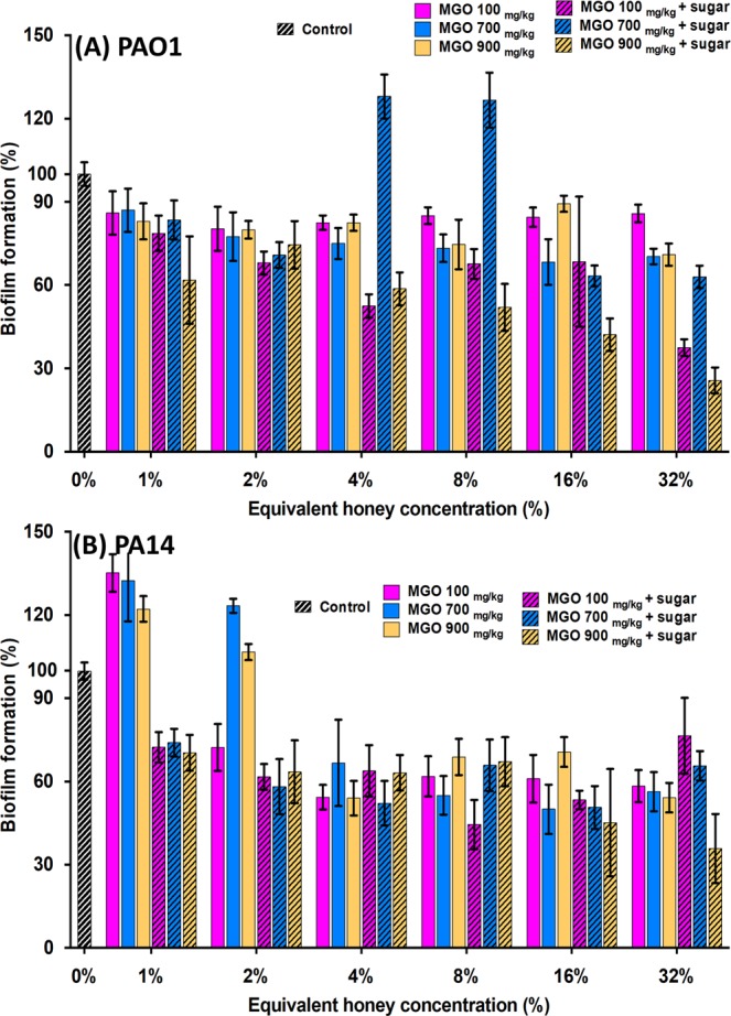
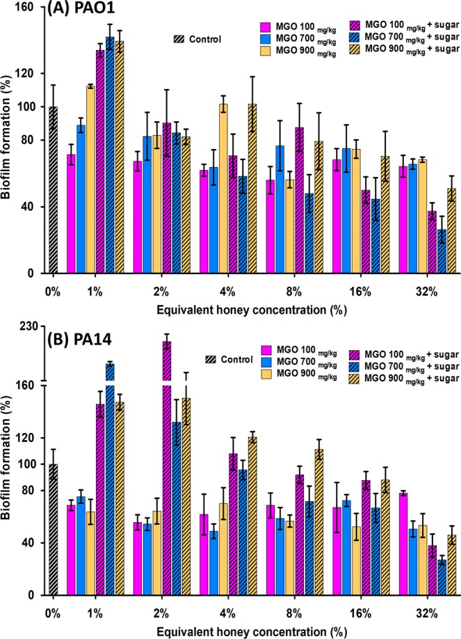
Chronic wound treatment is becoming increasingly difficult and costly, further exacerbated when wounds become infected. Bacterial biofilms cause most chronic wound infections and are notoriously resistant to antibiotic treatments. The need for new approaches to combat polymicrobial biofilms in chronic wounds combined with the growing antimicrobial resistance crisis means that honey is being revisited as a treatment option due to its broad-spectrum antimicrobial activity and low propensity for bacterial resistance. We assessed four well-characterised New Zealand honeys, quantified for their key antibacterial components, methylglyoxal, hydrogen peroxide and sugar, for their capacity to prevent and eradicate biofilms produced by the common wound pathogen Pseudomonas aeruginosa. We demonstrate that: (1) honey used at substantially lower concentrations compared to those found in honey-based wound dressings inhibited P. aeruginosa biofilm formation and significantly reduced established biofilms; (2) the anti-biofilm effect of honey was largely driven by its sugar component; (3) cells recovered from biofilms treated with sub-inhibitory honey concentrations had slightly increased tolerance to honey; and (4) honey used at clinically obtainable concentrations completely eradicated established P. aeruginosa biofilms. These results, together with their broad antimicrobial spectrum, demonstrate that manuka honey-based wound dressings are a promising treatment for infected chronic wounds, including those with P. aeruginosa biofilms.
Conflict of interest statement
Comvita New Zealand provided partial funding and materials (honey samples) for the work described in the manuscript. The authors declare that the research was conducted in the absence of any financial and non-financial relationships that could be construed as a potential conflict of interest.
Figures
Similar articles
-
Manuka-type honeys can eradicate biofilms produced by Staphylococcus aureus strains with different biofilm-forming abilities.PeerJ. 2014 Mar 25;2:e326. doi: 10.7717/peerj.326. eCollection 2014. PeerJ. 2014. PMID: 24711974 Free PMC article.
-
In vitro activity of an engineered honey, medical-grade honeys, and antimicrobial wound dressings against biofilm-producing clinical bacterial isolates.J Wound Care. 2016 Feb;25(2):93-4, 96-102. doi: 10.12968/jowc.2016.25.2.93. J Wound Care. 2016. PMID: 26878302
-
HOCl-producing electrochemical bandage is active in murine polymicrobial wound infection.Microbiol Spectr. 2024 Oct 3;12(10):e0062624. doi: 10.1128/spectrum.00626-24. Epub 2024 Aug 20. Microbiol Spectr. 2024. PMID: 39162542 Free PMC article.
-
Biofilms in Chronic Wound Infections: Innovative Antimicrobial Approaches Using the In Vitro Lubbock Chronic Wound Biofilm Model.Int J Mol Sci. 2023 Jan 5;24(2):1004. doi: 10.3390/ijms24021004. Int J Mol Sci. 2023. PMID: 36674518 Free PMC article. Review.
-
Evidence-Based Review of Antibiofilm Agents for Wound Care.Adv Wound Care (New Rochelle). 2021 Jan;10(1):13-23. doi: 10.1089/wound.2020.1193. Epub 2020 Jun 22. Adv Wound Care (New Rochelle). 2021. PMID: 32496980 Free PMC article. Review.
Cited by
-
Honey as an Ecological Reservoir of Antibacterial Compounds Produced by Antagonistic Microbial Interactions in Plant Nectars, Honey and Honey Bee.Antibiotics (Basel). 2021 May 9;10(5):551. doi: 10.3390/antibiotics10050551. Antibiotics (Basel). 2021. PMID: 34065141 Free PMC article. Review.
-
Advanced Wound Diagnostics: Toward Transforming Wound Care into Precision Medicine.Adv Wound Care (New Rochelle). 2022 Jun;11(6):330-359. doi: 10.1089/wound.2020.1319. Epub 2021 Jul 21. Adv Wound Care (New Rochelle). 2022. PMID: 34128387 Free PMC article. Review.
-
Evaluation of bacterial attachment on mineralized collagen scaffolds and addition of manuka honey to increase mesenchymal stem cell osteogenesis.Biomaterials. 2023 Mar;294:122015. doi: 10.1016/j.biomaterials.2023.122015. Epub 2023 Jan 19. Biomaterials. 2023. PMID: 36701999 Free PMC article.
-
Polyphenols Content and In Vitro α-Glycosidase Activity of Different Italian Monofloral Honeys, and Their Effect on Selected Pathogenic and Probiotic Bacteria.Microorganisms. 2021 Aug 9;9(8):1694. doi: 10.3390/microorganisms9081694. Microorganisms. 2021. PMID: 34442773 Free PMC article.
-
Comparison of the antimicrobial and antivirulence activities of Sidr and Tualang honeys with Manuka honey against Staphylococcus aureus.Iran J Microbiol. 2023 Feb;15(1):89-101. doi: 10.18502/ijm.v15i1.11923. Iran J Microbiol. 2023. PMID: 37069905 Free PMC article.
References
Publication types
MeSH terms
Substances
LinkOut - more resources
Full Text Sources
Medical
Molecular Biology Databases
