. 2019 Dec 3;9(1):18251.
doi: 10.1038/s41598-019-54683-0.

Elevated CO2 regulates the Wnt signaling pathway in mammals, Drosophila melanogaster and Caenorhabditis elegans

Affiliations

Elevated CO2 regulates the Wnt signaling pathway in mammals, Drosophila melanogaster and Caenorhabditis elegans

Masahiko Shigemura et al. Sci Rep. .

Abstract

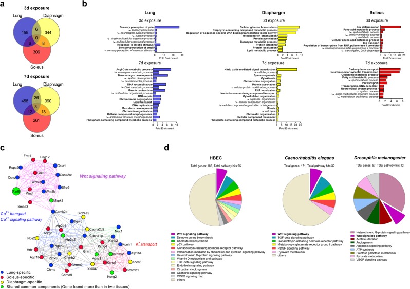
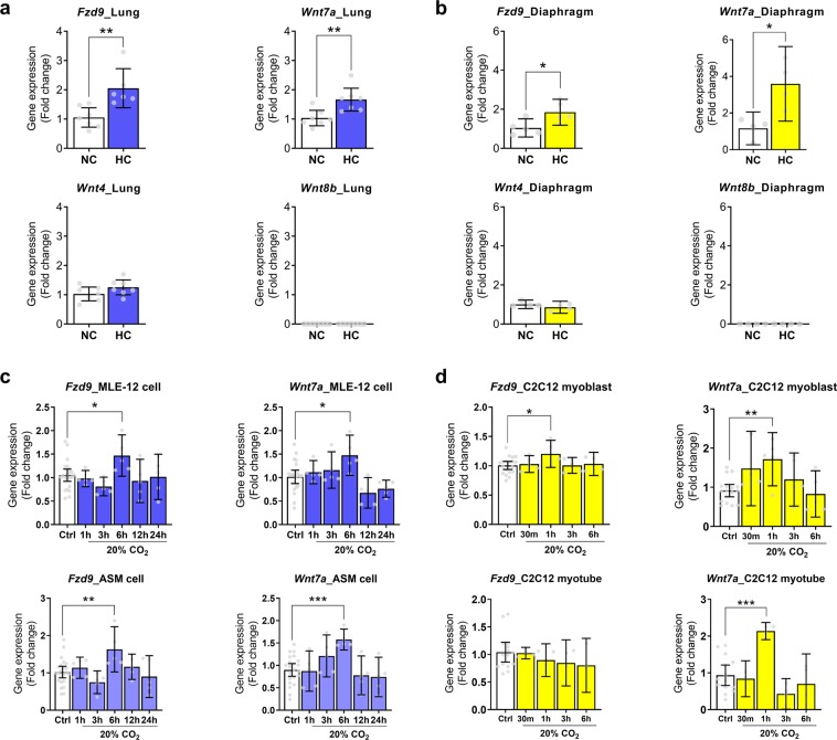
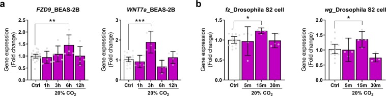
Carbon dioxide (CO2) is sensed by cells and can trigger signals to modify gene expression in different tissues leading to changes in organismal functions. Despite accumulating evidence that several pathways in various organisms are responsive to CO2 elevation (hypercapnia), it has yet to be elucidated how hypercapnia activates genes and signaling pathways, or whether they interact, are integrated, or are conserved across species. Here, we performed a large-scale transcriptomic study to explore the interaction/integration/conservation of hypercapnia-induced genomic responses in mammals (mice and humans) as well as invertebrates (Caenorhabditis elegans and Drosophila melanogaster). We found that hypercapnia activated genes that regulate Wnt signaling in mouse lungs and skeletal muscles in vivo and in several cell lines of different tissue origin. Hypercapnia-responsive Wnt pathway homologues were similarly observed in secondary analysis of available transcriptomic datasets of hypercapnia in a human bronchial cell line, flies and nematodes. Our data suggest the evolutionarily conserved role of high CO2 in regulating Wnt pathway genes.


Conflict of interest statement

The authors declare no competing interests.

Figures

Figure 1
Figure 1
A large-scale transcriptomic study of hypercapnia to combine mouse multi-tissue microarray analysis with secondary analysis of available transcriptomic datasets. (a–c) C57BL/6 J mice were exposed to normoxic hypercapnia for 3 (n = 4) or 7 (n = 3) days or maintained in sea-level room air (n = 3). (a) Venn diagrams showing the overlap between DEG from lung, diaphragm and soleus. (b) Gene ontology analysis of the DEG in each tissue using the PANTHER GO-Slim Biological Process annotation dataset. Arrows indicate hierarchical grouping between GO terms. (c) A network diagram constructed from the DEG in each dataset at 7-day exposure conditions. The network diagram revealed groups of genes and pathways that shared common components (green circles) but was also comprised of lung-specific, diaphragm-specific, soleus-specific responsive to hypercapnic exposure (blue, yellow and red circles, respectively). (d) Secondary analysis of transcriptomic datasets of hypercapnia in HBEC, Caenorhabditis elegans and Drosophila melanogaster. PANTHER classification system categorized the DEG in each dataset into signaling pathways.
Figure 2
Figure 2
Validation of mouse multi-tissue microarray analysis. (a,b) Validation in mouse tissues. Fzd9, Wnt4, Wnt7a, and Wnt8b expression in the lung (n = 6–7) (a) and diaphragm skeletal muscle (n = 4–5) (b) from mice exposed to normoxic hypercapnia for 7 days. NC, normocapnia; HC, hypercapnia. (c,d) Validation in mouse lung and skeletal muscle cells. Fzd9 and Wnt7a expressions in MLE-12 cells (Ctrl, n = 22–23; 20%CO2, n = 5 per group), ASM cells (Ctrl, n = 22–23; 20%CO2, n = 5 per group) (c), or C2C12 myoblast (Ctrl, n = 18–19; 20%CO2, n = 4–5 per group) or myotube (Ctrl, n = 14–15; 20%CO2, n = 3–4 per group) (d) exposed to high CO2 for up to 24 hours (c) or 6 hours (d). Ctrl, control conditions. All values are represented as mean with error bars shown as the 95% confidence interval. *p < 0.05, **p < 0.01, ***p < 0.001, unpaired two-tailed Student’s t test or one-way ANOVA with Dunnett’s post hoc test.
Figure 3
Figure 3
Validation of the transcriptomic datasets of hypercapnia in a human bronchial cell line and invertebrates. (a) FZD9 and WNT7a expression in BEAS-2B cells exposed to high CO2 for up to 12 hours (Ctrl, n = 20–23; 20%CO2, n = 5–6 per group). Ctrl, control conditions. (b) Fz and wg expression in Drosophila S2 cells exposed to high CO2 for up to 30 min (Ctrl, n = 14–15; 20%CO2, n = 4–5 per group). All values are represented as mean with error bars shown as the 95% confidence interval. *p < 0.05, **p < 0.01, ***p < 0.001, one-way ANOVA with Dunnett’s post hoc test.

References

    1. Weir EK, Lopez-Barneo J, Buckler KJ, Archer SL. Acute oxygen-sensing mechanisms. N. Engl. J. Med. 2005;353:2042–2055. doi: 10.1056/NEJMra050002. - DOI - PMC - PubMed
    1. Haldar SM, Stamler JS. S-nitrosylation: integrator of cardiovascular performance and oxygen delivery. J. Clin. Invest. 2013;123:101–110. doi: 10.1172/JCI62854. - DOI - PMC - PubMed
    1. Cummins EP, Selfridge AC, Sporn PH, Sznajder JI, Taylor CT. Carbon dioxide-sensing in organisms and its implications for human disease. Cell. Mol. Life Sci. 2014;71:831–845. doi: 10.1007/s00018-013-1470-6. - DOI - PMC - PubMed
    1. Shigemura M, Lecuona E, Sznajder JI. Effects of hypercapnia on the lung. J. Physiol. 2017;595:2431–2437. doi: 10.1113/JP273781. - DOI - PMC - PubMed
    1. Laffey JG, Kavanagh BP. Carbon dioxide and the critically ill–too little of a good thing? Lancet. 1999;354:1283–1286. doi: 10.1016/S0140-6736(99)02388-0. - DOI - PubMed

Publication types

LinkOut - more resources