

Conjunctival pigmented lesion: Clinicopathological analysis of 85 cases in Korean population
- PMID: 31796811
- PMCID: PMC6890774
- DOI: 10.1038/s41598-019-54786-8
Conjunctival pigmented lesion: Clinicopathological analysis of 85 cases in Korean population
Abstract
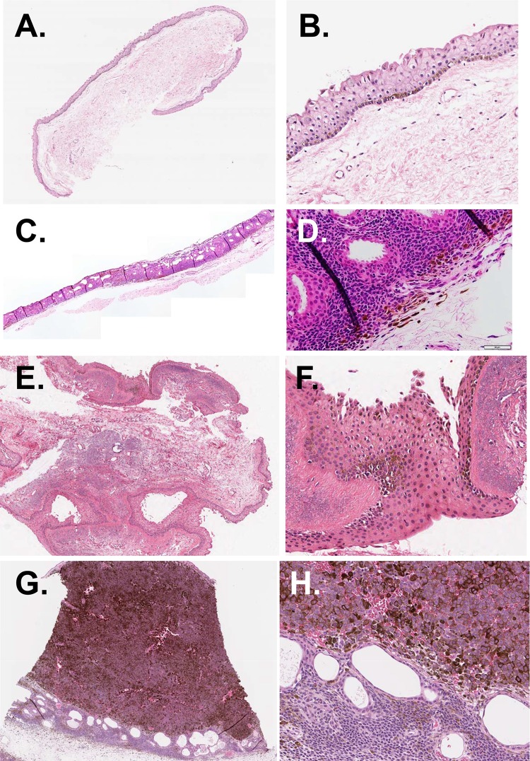
To evaluate histopathological characteristics of conjunctival pigmented lesions and analyze clinical features related to histologic classification in Asian population, we analyzed medical records, anterior segment photographs, and histological specimen of 85 eyes who had undergone biopsy for pigmented conjunctival lesions at Seoul National University Hospital between 1999 and 2018. Compound nevus was the most common type of conjunctival pigmented lesions (67.1%), followed by conjunctival melanocytic intraepithelial neoplasia (primary acquired melanosis)(11.8%), subepithelial nevus (8.2%), and malignant melanoma (MM)(7.1%). Patients with compound nevus were younger than those with non-compound nevus (22.1 ± 17.0 vs 39.9 ± 18.8 years, p < 0.001), while patients with MM were older than those without melanoma (55.7 ± 18.2 vs 25.8 ± 18.0 years, p = 0.001). The lesion in compound nevus tended to be more frequently located on the temporal conjunctiva than that in the non-compound nevus group (54.4% vs 32.1%, p = 0.053), and feeder vessels were associated with most of compound nevus (98.2% vs 78.6% of non-compound nevus, p = 0.005). The lesion in MM was larger, involved multiple quadrants, and had extrabulbar location than lesions without melanoma (p < 0.001, p < 0.001, and p = 0.002, respectively). Together, the results would help clinicians to distinguish benign conjunctival pigmentations from malignant counterparts in clinical practice without biopsy.
Conflict of interest statement
The authors declare no competing interests.
Figures

References
-
- Zembowicz A, Mandal RV, Choopong P. Melanocytic lesions of the conjunctiva. Arch. Pathol. Lab. Med. 2010;134:1785–1792. - PubMed
MeSH terms
LinkOut - more resources
Full Text Sources
Medical
