

Adaptive weighted log subtraction based on neural networks for markerless tumor tracking using dual-energy fluoroscopy
- PMID: 31797397
- PMCID: PMC7015793
- DOI: 10.1002/mp.13941
Adaptive weighted log subtraction based on neural networks for markerless tumor tracking using dual-energy fluoroscopy
Abstract
Purpose: To present a novel method, based on convolutional neural networks (CNN), to automate weighted log subtraction (WLS) for dual-energy (DE) fluoroscopy to be used in conjunction with markerless tumor tracking (MTT).
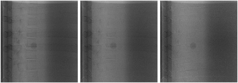
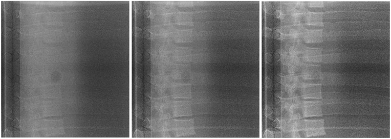
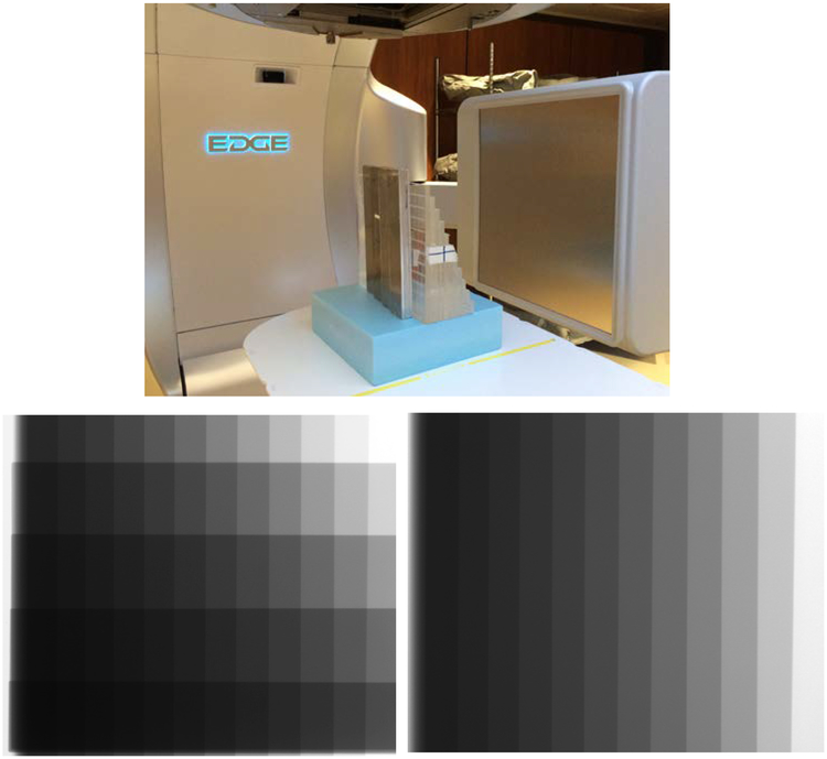
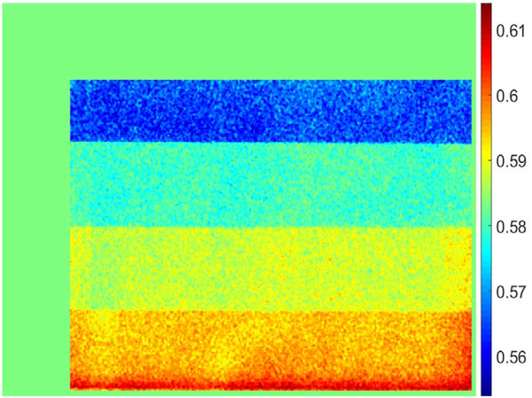
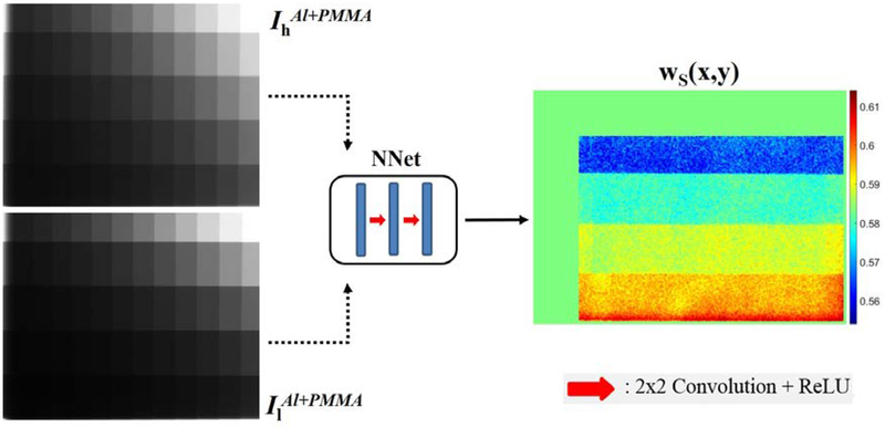
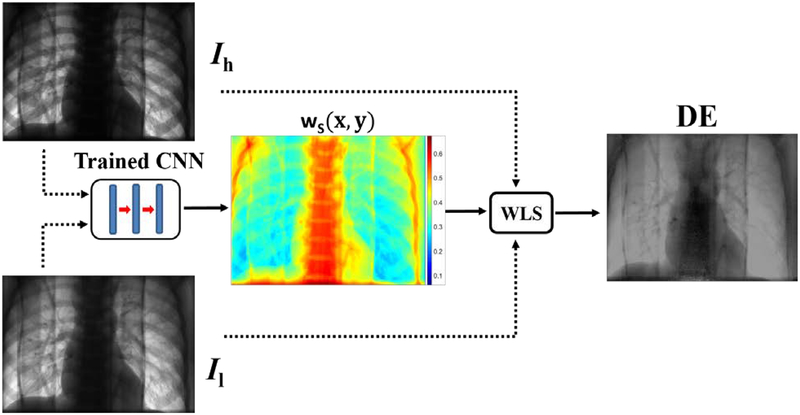
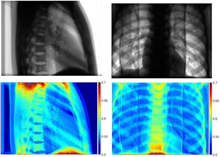
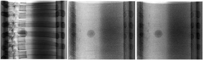
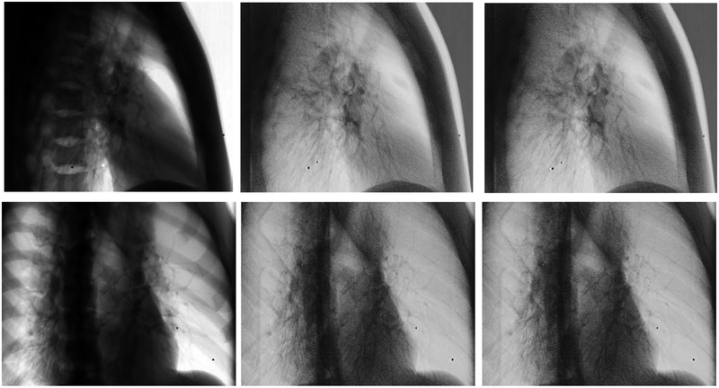
Methods: A CNN was developed to automate WLS (aWLS) of DE fluoroscopy to enhance soft tissue visibility. Briefly, this algorithm consists of two phases: training a CNN architecture to predict pixel-wise weighting factors followed by application of WLS subtraction to reduce anatomical noise. To train the CNN, a custom phantom was built consisting of aluminum (Al) and acrylic (PMMA) step wedges. Per-pixel ground truth (GT) weighting factors were calculated by minimizing the contrast of Al in the step wedge phantom to train the CNN. The pretrained model was then utilized to predict pixel-wise weighting factors for use in WLS. For comparison, the weighting factor was manually determined in each projection (mWLS). A thorax phantom with five simulated spherical targets (5-25 mm) embedded in a lung cavity, was utilized to assess aWLS performance. The phantom was imaged with fast-kV dual-energy (120 and 60 kVp) fluoroscopy using the on-board imager of a commercial linear accelerator. DE images were processed offline to produce soft tissue images using both WLS methods. MTT was compared using soft tissue images produced with both mWLS and aWLS techniques.
Results: Qualitative evaluation demonstrated that both methods achieved soft tissue images with similar quality. The use of aWLS increased the number of tracked frames by 1-5% compared to mWLS, with the largest increase observed for the smallest simulated tumors. The tracking errors for both methods produced agreement to within 0.1 mm.
Conclusions: A novel method to perform automated WLS for DE fluoroscopy was developed. Having similar soft tissue quality as well as bone suppression capability as mWLS, this method allows for real-time processing of DE images for MTT.
Keywords: convolutional neural networks; dual-energy imaging; fast-kV switching; markerless tumor tracking.
© 2019 American Association of Physicists in Medicine.
Figures
References
-
- Brody WR, Butt G, Hall A, and Macovski A, A method for selective tissue and bone visualization using dual energy scanned projection radiography, Med. Phys 8(3), 353–357 (1981). - PubMed
-
- Shkumat NA, Siewerdsen JH, Richard S, Paul NS, Yorkston J, and Van Metter R, Dual-energy imaging of the chest: Optimization of image acquisition techniques for the “bone-only” image, Med. Phys 35(2), 629–632 (2008). - PubMed
-
- Block AM, Patel R, Panfil J, et al. , Evaluation of a Template-Based Algorithm for Markerless Lung Tumor Tracking on Single Energy and Dual Energy kV Images, Int. J. Radiat. Oncol 90(1), S142–S143 (2014).
-
- Patel R, Panfil J, Campana M, et al. , Markerless motion tracking of lung tumors using dual-energy fluoroscopy, Med. Phys 42(1), 254–262 (2015). - PubMed
-
- Hoggarth MA, Luce J, Syeda F, et al. , Dual energy imaging using a clinical on-board imaging system, Phys. Med. Biol 58(12), 4331–4340 (2013). - PubMed
MeSH terms
Grants and funding
LinkOut - more resources
Full Text Sources
Medical
Miscellaneous
