. 2021 Jun;18(6):1463-1475.
doi: 10.1038/s41423-019-0331-0. Epub 2019 Dec 4.

Serum-circulating His-tRNA synthetase inhibits organ-targeted immune responses

Affiliations

Serum-circulating His-tRNA synthetase inhibits organ-targeted immune responses

Ryan A Adams et al. Cell Mol Immunol. 2021 Jun.

Abstract

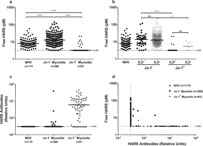
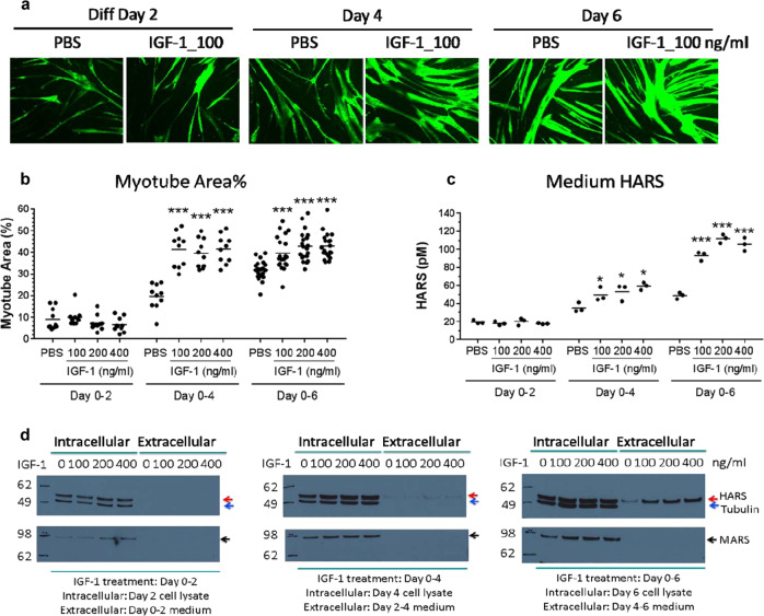
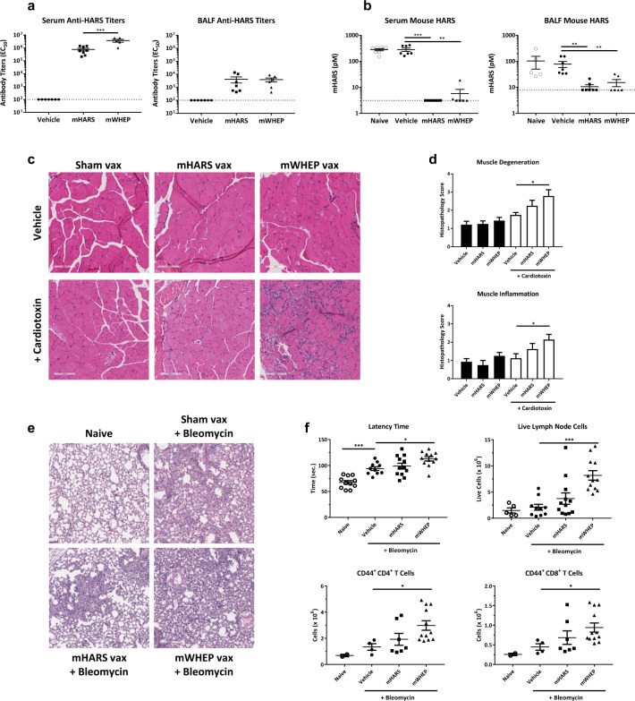
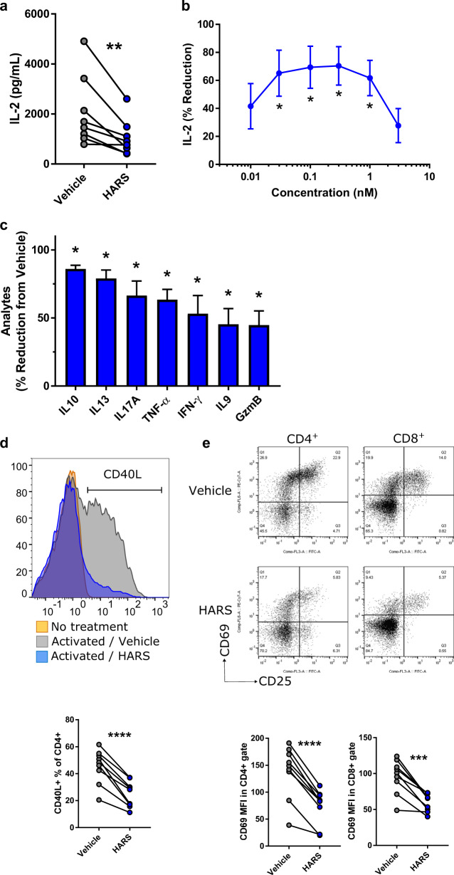
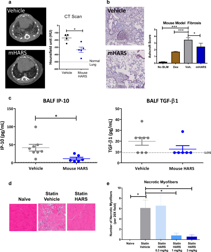
His-tRNA synthetase (HARS) is targeted by autoantibodies in chronic and acute inflammatory anti-Jo-1-positive antisynthetase syndrome. The extensive activation and migration of immune cells into lung and muscle are associated with interstitial lung disease, myositis, and morbidity. It is unknown whether the sequestration of HARS is an epiphenomenon or plays a causal role in the disease. Here, we show that HARS circulates in healthy individuals, but it is largely undetectable in the serum of anti-Jo-1-positive antisynthetase syndrome patients. In cultured primary human skeletal muscle myoblasts (HSkMC), HARS is released in increasing amounts during their differentiation into myotubes. We further show that HARS regulates immune cell engagement and inhibits CD4+ and CD8+ T-cell activation. In mouse and rodent models of acute inflammatory diseases, HARS administration downregulates immune activation. In contrast, neutralization of extracellular HARS by high-titer antibody responses during tissue injury increases susceptibility to immune attack, similar to what is seen in humans with anti-Jo-1-positive disease. Collectively, these data suggest that extracellular HARS is homeostatic in normal subjects, and its sequestration contributes to the morbidity of the anti-Jo-1-positive antisynthetase syndrome.

Keywords: Autoimmunity; HARS; Immunology; Synthetase; tRNA.


Conflict of interest statement

The authors declare no competing interests.

Figures

Fig. 1
Fig. 1. Free HARS is present in the circulation and low or absent in anti-Jo-1-positive disease.
a Free-serum HARS levels measured in normal human volunteers (n = 115), anti-Jo-1-positive myositis patients (n = 61), and patients with anti-Jo-1-negative myositis (n = 286). Each dot represents an individual patient. Bars represent median values. Significantly lower levels of free HARS (****p < 0.0001) were observed in the anti-Jo-1-positive myositis patients compared to normal individuals and anti-Jo-1-negative myositis (n = 286) patients. b Serum HARS level in patients with ILD (n = 94) and without ILD (n = 243) were segmented by Jo-1 status (ILD+ Jo-1 n = 50, ILD Jo-1 n = 236, ILD+, Jo-1+ n = 44, ILD Jo-1+ n = 7). c HARS-specific antibody levels in healthy volunteers were compared to individuals with Jo-1-positive and Jo-1-negative myositis (sample numbers as in a). d Correlation of HARS serum levels with anti-HARS antibodies (data from panels a and c).
Fig. 2
Fig. 2. IGF-1 stimulation enhanced myotube differentiation and HARS release from HSkMC.
a HSkMC were treated with IGF-1 or PBS during differentiation days 0–2, 0–4, or 0–6 with medium renewal every 2 days. Cells were fixed on differentiation days 2, 4, or 6 for staining (representative images shown) and analysis of myotube area (panel b). c HARS in the medium was accumulated for the last two days before harvest and was quantified by ELISA. The results are shown as the mean ± SEM. For myotube area, fusion index and nuclei number, n = 20 images. For HARS release, n = 3 biological replicates. The p-values of <0.05 (*) or 0.001 (***) are indicated. d Western blot analysis showed an increase in extracellular HARS but not MARS or Tubulin proteins in the HSkMC medium upon IGF-1 treatment.
Fig. 3
Fig. 3. Antibodies specific to the immunomodulatory domain in mice mimic some features of human anti-Jo-1-positive antisynthetase syndrome.
a Anti-HARS antibody titers in serum (left panel) and BALF (right panel) were measured by ECLIA. Immunized animals were all titer positive. The dotted line represents the limit of quantitation, and the bars represent the mean ± SEM. b Serum and BALF HARS were measured by ECLIA in control animals (naive), vehicle with control antigen, mouse HARS (mHARS), or mouse HARS WHEP domain (mWHEP). Serum HARS levels were reduced in both sets of immunizations (***p < 0.005, **p < 0.01; one-way ANOVA). BALF HARS levels were suppressed in immunized animals (**p < 0.01; one-way ANOVA). c Representative images of the right soleus from mice subjected to vehicle treatment (Sham Vax), mouse HARS or mouse HARS WHEP immunization followed by intramuscular (right tibialis anterior, gastrocnemius and quadriceps) injection with vehicle or cardiotoxin. Note the lack of immune cell infiltration in animals immunized with HARS WHEP in the absence of cardiotoxin, and also note the exacerbated inflammatory infiltrate in animals receiving cardiotoxin in the presence of antibodies against the HARS WHEP domain. x20 magnification. d Quantification of soleus muscle degeneration and inflammation based on the following semiquantitative scoring system: 0 = no significant lesion; minimal change = 1; mild change = 2; moderate change = 3; and marked change = 4. A score of 1 may, and often does, represent an incidental lesion that could be found randomly in normal animals. *p < 0.05, one-way ANOVA. e Representative H&E images of the lungs from animals that were naive or subjected to control immunization (Sham Vax), mHARS, or mWHEP immunization, and were then challenged with bleomycin. x10 magnification. f Upper left panel: the latency time required to become anesthetized by the inhaled anesthetic isoflurane was lengthened in animals challenged with bleomycin, suggesting impaired gas exchange in these animals. The latency time was even greater in animals immunized with mWHEP prior to bleomycin challenge. Upper right panel: the number of live cells isolated from the mediastinal lymph nodes of mice immunized with mWHEP and challenged with bleomycin is greater than the number present in sham-immunized bleomycin-challenged animals. Lower panels: the number of activated CD4 + (left) and CD8 + T cells (right) isolated from the mediastinal lymph nodes of mice immunized with mWHEP and challenged with bleomycin is greater than the number present in sham-immunized bleomycin-challenged animals. *p < 0.05, ***p < 0.001 one-way ANOVA followed by Dunnett’s post hoc vs. Vehicle vax/BLM challenged group.
Fig. 4
Fig. 4. T-cell activation is reduced by HARS-related proteins.
T cells isolated from PBMCs obtained from healthy volunteers were stimulated with plate-bound anti-CD3/anti-CD28 for 24 h in the presence of vehicle or recombinant human HARS.a IL-2 release by individual donors (n = 8) was measured, [HARS] = 0.3 nM. b IL-2 reduction by treatment with the vehicle control (n = 3) was measured after treatment with HARS at different concentrations. c Effect of HARS (0.3 nM) on the release of various cytokines and granzyme B (n = 2 donors in two separate experiments) were measured. d Flow cytometry analysis of CD40L expression on CD4 + T cells was performed, and the representative histogram and % positive results from individual donors (n = 9) are shown, [HARS] = 1 nM. e Flow cytometry analysis of CD69 expression on CD4 + (left) and CD8 + (right) T cells was performed, and representative scatter plots and MFI results from individual donors (n = 9) are shown, [HARS] = 1 nM. Error bars indicate SEM, paired t-test or ANOVA with Dunnett’s post hoc test where appropriate. *p < 0.05; **p < 0.01; ***p < 0.001, ****p < 0.0001.
Fig. 5
Fig. 5. Addition of HARS can ameliorate immune-driven disease.
a Left panel: Anatomically matched representative CT images from mice 14 days after challenge with bleomycin and treatment for 7 days with vehicle or mouse HARS. Right panel: CT scans were quantified by a mean of eight regions of interest per animal using Hounsfield units (HU). Vehicle-treated animals had an elevated mean Hounsfield unit, whereas HARS decreased the values toward those of the normal lung. b Representative H&E stained sections from bleomycin-induced mice terminated 21 days after challenge with bleomycin and treatment with vehicle or mouse HARS beginning on day 8. Note the immune infiltrate, fibrosis and lack of open alveoli in vehicle-treated animals and improved histology in mice treated with HARS. Right panel: Fibrosis scored on a modified Ashcroft scale in mice challenged with bleomycin on Day 0 and treated as indicated starting on Day 0 (dex) or starting on Day 8 (vehicle IV, mHARS). c BALF levels of IP-10 and TGFβ1 were measured from mice represented in panel b. d In a moderate statin-induced myositis model, representative histological images of hamstrings were obtained on Day 15 from animals naive to statin or receiving statin plus vehicle or statin plus HARS (3 mg/kg) beginning on Day 6 following statin initiation. e Necrotic fibers in statin-treated mice were counted by a person blinded to the treatment groups. HARS was administered at 0.3, 1, or 3 mg/kg on days 6–14, and muscle analysis was conducted on day 15. *p < 0.01, one-way ANOVA followed by Dunnett’s post hoc test.

References

    1. Mahler M, Miller FW, Fritzler MJ. Idiopathic inflammatory myopathies and the anti-synthetase syndrome: a comprehensive review. Autoimmmun. Rev. 2014;13:367–371. - PMC - PubMed
    1. Hervier B, Benveniste O. Clinical heterogeneity and outcomes of antisynthetase syndrome. Curr. Rheumatol. Rep. 2013;15:349. - PubMed
    1. Mathews MB, Bernstein RM. Myositis autoantibody inhibits histidyl-tRNA synthetase: a model for autoimmunity. Nature. 1983;304:177–179. - PubMed
    1. Ramsden DA, et al. Epitope mapping of the cloned human autoantigen, histidyl-tRNA synthetase. Analysis of the myositis-associated anti-Jo-1 autoimmune response. J. Immunol. 1989;143:2267–2272. - PubMed
    1. Amato AA, Greenberg SA. Inflammatory myopathies. CONTINUUM: Lifelong Learn. Neurol. 2013;19:1598–1614. - PMC - PubMed

Publication types

MeSH terms

Supplementary concepts