. 2019 Dec 4;10(1):5527.
doi: 10.1038/s41467-019-13440-7.

Self-regenerating giant hyaluronan polymer brushes

Affiliations

Self-regenerating giant hyaluronan polymer brushes

Wenbin Wei et al. Nat Commun. .

Abstract

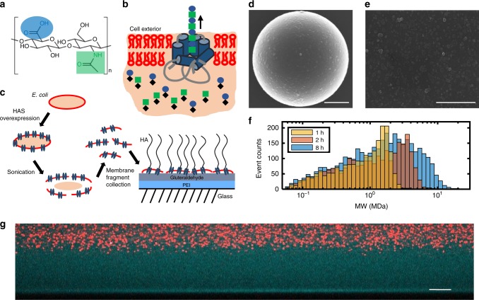
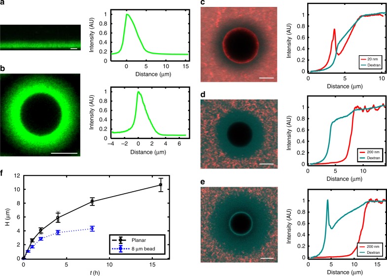
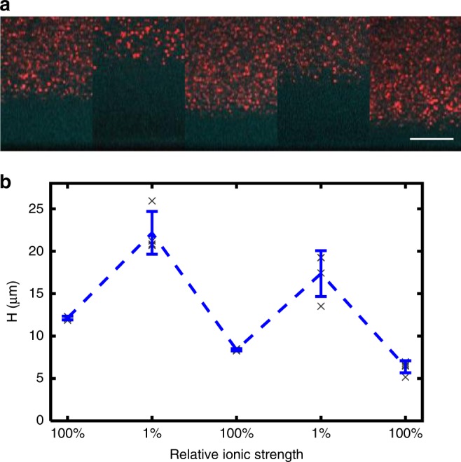
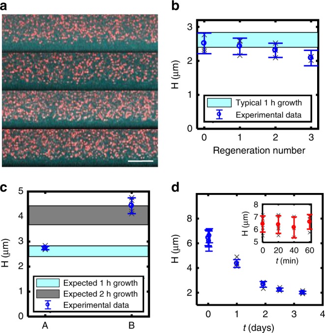
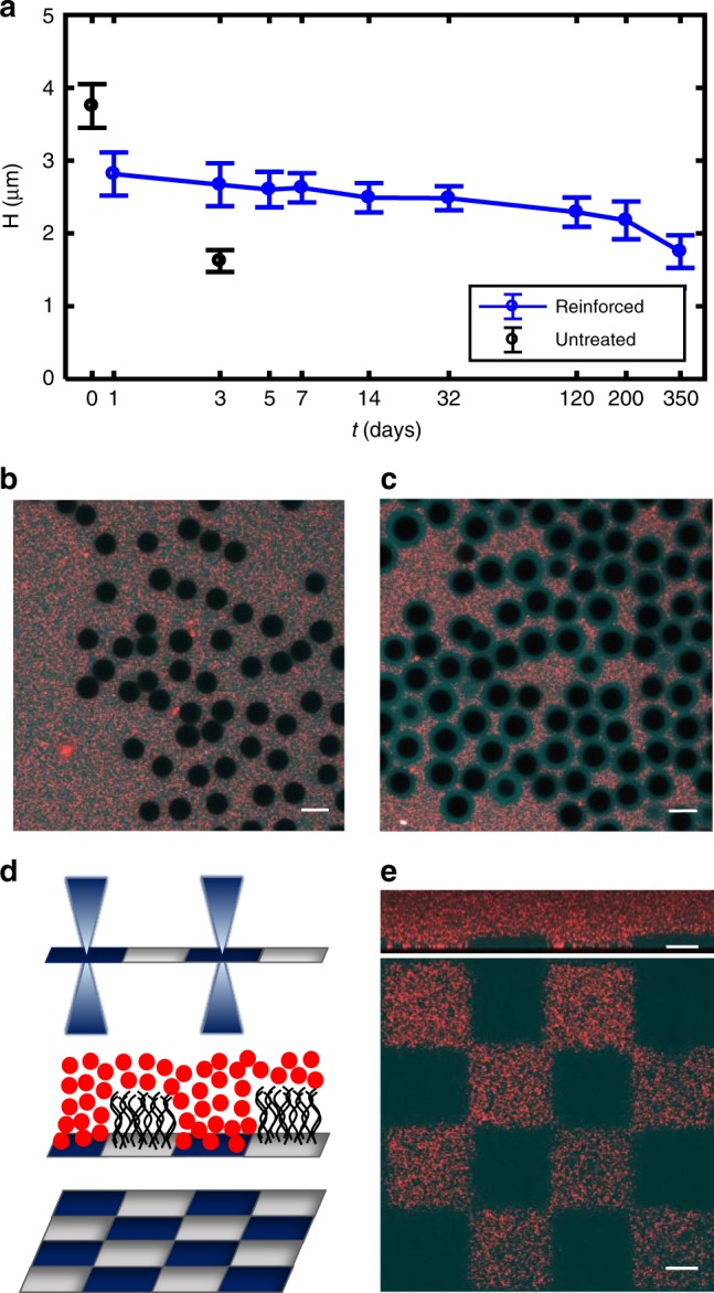
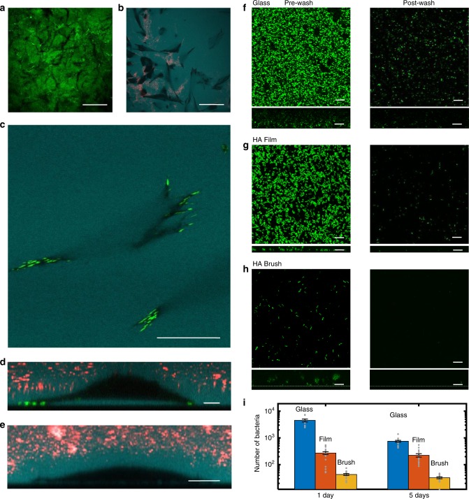
Tailoring interfaces with polymer brushes is a commonly used strategy to create functional materials for numerous applications. Existing methods are limited in brush thickness, the ability to generate high-density brushes of biopolymers, and the potential for regeneration. Here we introduce a scheme to synthesize ultra-thick regenerating hyaluronan polymer brushes using hyaluronan synthase. The platform provides a dynamic interface with tunable brush heights that extend up to 20 microns - two orders of magnitude thicker than standard brushes. The brushes are easily sculpted into micropatterned landscapes by photo-deactivation of the enzyme. Further, they provide a continuous source of megadalton hyaluronan or they can be covalently-stabilized to the surface. Stabilized brushes exhibit superb resistance to biofilms, yet are locally digested by fibroblasts. This brush technology provides opportunities in a range of arenas including regenerating tailorable biointerfaces for implants, wound healing or lubrication as well as fundamental studies of the glycocalyx and polymer physics.


Conflict of interest statement

The authors declare no competing interests.

Figures

Fig. 1
Fig. 1
Construction of the hyaluronan synthase brush. a The disaccharide unit of HA is comprised of d-glucuronic acid (GlcUA) and N-acetyl-d-glucosamine (GlcNAc). b HA synthase (blue) embedded in a bacterial membrane (red) polymerizes and extrudes the growing HA polymer through the pore. The sugar substrates UDP-GlcUA (circle) and UDP-GlcNAc (square) are alternatively bound at the intracellular glycotransferase sites where processive assembly takes place. HA is extruded at an average rate of ~1 nm/s corresponding to 1 disaccharide/s. c Preparation and immobilization of membrane fragments carrying HA synthase to glass substrates. d, e SEM images of the membrane fragments on spherical and planar surfaces. Scale bars 2 µm. f HA molecular weight distributions assayed by solid-state nanopore (N = 2091, 1 h; N = 2500, 2 h; N = 3699, 8 h). See Supplementary Info, Table 1. g Side view confocal image of HA brush grown for 16 h and imaged at low ionic strength (1.5 mM). The brush region is imaged by the contrast generated by its accessibility to fluorescent dextran (cyan, 10 kDa), but exclusion of nanoparticles (red, 200 nm). The brush height is ~22 µm. Scale bar 10 µm.
Fig. 2
Fig. 2
Brush concentration profiles, nanoparticle sieving and growth dynamics. a Fluorescent profile of a planar HA brush and its intensity profile. b Fluorescent profile of spherical brush and its intensity profile. In both a, b the brush growth time is 4 h at 30 °C and the ionic strength 150 mM. ce Cross-section of brush grown for 4 h on 8 µm microsphere (left) and the particle penetration intensity profile (right) for c 20 nm, 150 mM, d 200 nm, 150 mM and e 200 nm, 1.5 mM. Twenty nanometer particles show a distinct gradient within the brush while 200 nm particles remain excluded. Dextran (10 kDa, ~5 nm) is present in all cases (cyan) and shows an enhanced gradient in the low ionic strength swollen brush. f Dynamic growth of hyaluronan brushes generated by HA synthase (150 mM). The brush height reaches 2.62 ± 0.2 µm (st. dev.) in just 1 h (planar) in high ionic strength conditions. After 16 h the brush is 10.6 ± 1.0 µm (st. dev.). Measurements from individual brushes are shown in gray, averages and standard deviations are indicated in black. N_planar = 3 brushes, 12 regions per sample, except 16 h brush which is just one brush. The spherical brush plateaus at much earlier times (~5 h) at a final height of 4.3 ± 0.4 µm (st. dev.). N_spherical > 120 for spherical brush height measurements. All scale bars are 5 µm.
Fig. 3
Fig. 3
Stimulus responsiveness and reversibility. a Brush height during a series of solvent swaps from 133 to 1.33 mM for a brush previously grown for 16 h. Scale bar, 10 µm. b Quantification of the brush height shows that at ultra-low ionic strengths, the brush stretches out by nearly 200%, peaking at 22.0 ± 2.5 µm (st. dev.) during the first exchange. While the brush swelling and shrinking is reversible, the repeated handling (and tension induced by stretching) leads to some loss of the HA, which is weakly bound to the HA synthase. As a consequence, a gradual decrease in the overall brush height is observed. Each gray data point corresponds to five independent measurements (211 × 211 µm2 area) from one sample. Blue data points show the mean and st. dev.
Fig. 4
Fig. 4
Brush regeneration and patterning. a Regeneration of HA brush after enzymatic degradation with hyaluronidase. Top image shows brush after 1 h of growth before digestion. The next three images show the regenerated brush following digestion and 1 h regrowth 1, 2, and 3 times. b Brush height versus the number of regeneration times (N = 3 brushes, where gray x’s correspond to average of five measurements of each brush and blue is the mean and st dev). c Interrupted growth (A) followed by an additional growth period of 1 h (B). (Gray x’s correspond to five measurements from one sample.) d Brush stability versus time (unreinforced, natural brush), For both c, d, N = 1 brush, gray x’s correspond to measurements on same brush, blue reports the mean and st. dev.
Fig. 5
Fig. 5
Reinforcement stabilizes HA brush. a Reinforced spherical brush height measured over a period of 1 year versus natural, untreated spherical brush height. For each time point the corresponding number of measurements are N = 35, 126, 119, 103, 64, 56, 62, 76, 59, 28. Error bars report st. dev. b Reinforced brushes on spherical particles are still digested by hyaluronidase, indicating minimal HA−HA crosslinking within the brush. c Reinforced brushes on spherical particles resist detergent treatment (SDS) despite the removal of membrane fragments. This confirms the HA is stably bound to the underlying glass substrate. d UV micropatterning of HA synthase activity with a confocal microscope (λ = 405 nm). e Checkerboard pattern illustrating binary patterning of the brush. Red regions indicate areas where no brush grew; cyan regions indicate regions where brush expels red nanoparticles. Top: XZ confocal side view of micropatterned brush. Bottom: XY confocal image taken at the glass interface. All data in this figure were acquired in physiological conditions (150 mM). All scale bars are 10 µm.
Fig. 6
Fig. 6
Fibroblast and biofilm interactions with HA brush. a Black tracks in the GFPn-labeled HA brush reveal areas of brush digestion by MEF cells. Scale bar 50 µm. b Fluorescent dextran (cyan) highlights space under adherent cells (to the brush). Black regions correspond to cell area in close contact with surface. Red fluorescent particles fill areas of brush elimination due to digestion by cells. Scale bar 50 µm. c Fibroblasts attach to the interface underlying the HA brush, expressing mature focal adhesions (green, vinculin). Dextran is apparent underneath the spread cell. Scale bar 20 µm. d z-slice of the same adherent fibroblast from (c) sitting on a cushion of HA. Scale bar 10 µm. e Particle exclusion assay reveals MEFs express a thick glycocalyx. All MEF images were taken after 12 h exposure to the HA reinforced brush. Scale bar 10 µm. fh Confocal micrographs of GFP-producing Pseudomonas aeruginosa (PAO1) interacting with a glass interface (f), an HA film (g), and a reinforced HA brush (h). All images were taken at the glass interface. Left: biofilm growth before washing (1 day). Right: biofilm growth after washing (1 day). Dextran was used to identify the glass interface beneath the brush. XZ side views of the biofilms are presented below each respective XY top view of the samples. Scale bars, 10 µm in (fh). i Comparison of the number of bacteria retained after washing different surfaces. Data were taken in triplicates and averaged over five regions per sample. Error is SEM. Data are available in Table 3 in the Supplementary Information.

References

    1. Webber MJ, Appel EA, Meijer EW, Langer R. Supramolecular biomaterials. Nat. Mater. 2015;15:13. doi: 10.1038/nmat4474. - DOI - PubMed
    1. Burdick JA, Murphy WL. Moving from static to dynamic complexity in hydrogel design. Nat. Commun. 2012;3:1269. doi: 10.1038/ncomms2271. - DOI - PubMed
    1. Rosales AM, Anseth KS. The design of reversible hydrogels to capture extracellular matrix dynamics. Nat. Rev. Mater. 2016;1:15012. doi: 10.1038/natrevmats.2015.12. - DOI - PMC - PubMed
    1. Ahn BK, Lee DW, Israelachvili JN, Waite JH. Surface-initiated self-healing of polymers in aqueous media. Nat. Mater. 2014;13:867. doi: 10.1038/nmat4037. - DOI - PubMed
    1. DeForest CA, Anseth KS. Cytocompatible click-based hydrogels with dynamically tunable properties through orthogonal photoconjugation and photocleavage reactions. Nat. Chem. 2011;3:925. doi: 10.1038/nchem.1174. - DOI - PMC - PubMed

Publication types