. 2019 Dec 4;10(1):5531.
doi: 10.1038/s41467-019-13438-1.

CD4+ T cell help creates memory CD8+ T cells with innate and help-independent recall capacities

Affiliations

CD4+ T cell help creates memory CD8+ T cells with innate and help-independent recall capacities

Tomasz Ahrends et al. Nat Commun. .

Abstract

CD4+ T cell help is required for the generation of CD8+ cytotoxic T lymphocyte (CTL) memory. Here, we use genome-wide analyses to show how CD4+ T cell help delivered during priming promotes memory differentiation of CTLs. Help signals enhance IL-15-dependent maintenance of central memory T (TCM) cells. More importantly, help signals regulate the size and function of the effector memory T (TEM) cell pool. Helped TEM cells produce Granzyme B and IFNγ upon antigen-independent, innate-like recall by IL-12 and IL-18. In addition, helped memory CTLs express the effector program characteristic of helped primary CTLs upon recall with MHC class I-restricted antigens, likely due to epigenetic imprinting and sustained mRNA expression of effector genes. Our data thus indicate that during priming, CD4+ T cell help optimizes CTL memory by creating TEM cells with innate and help-independent antigen-specific recall capacities.


Conflict of interest statement

The authors declare no competing interests.

Figures
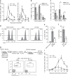
Fig. 1
Fig. 1
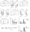
CD4+ T cell help instills intrinsic recall capacities into CD8+ T cells. af Mice were vaccinated intra-epidermally with a DNA construct encoding HPV-E7 with (Help) or without (No Help) MHC class II-restricted epitopes on days 0, 3, and 6. On day 50, mice were rechallenged with No Help vaccine and i.p. lipopolysaccharide (LPS) injection. (A) Percentage of H-2Db/E749-57 tetramer+ cells among total CD8+ T cells in blood at indicated days after first vaccination (n = 7 per group). b, c In a separate experiment, on day 10 after rechallenge, frequencies of Granzyme B (GZMB)+ cells (B) and IFNγ+TNFα+ cells (C) within total CD8+ T cells in indicated tissues were determined by flow cytometry (n = 3–4). d, e In a separate experiment, mice (n = 3) were injected i.v on day 10 after rechallenge with a 1:1 ratio of splenocytes loaded with E749-57 peptide (CTL target) or not (control) and labeled with a low or high dose of carboxyfluorescein succinimidyl ester (CFSE), respectively. Naive recipient mice were used as control. After 15 h, percentage of specific in vivo target cell killing was determined by flow cytometric analysis of splenocytes. d Representative histograms of CFSE+ cells. e Bar diagram quantitatively depicting all results. f In a separate experiment, E7-specific CD8+ T cells were isolated from spleen at day 10 after rechallenge (n = 3). T cells were incubated in vitro at a 1:1 ratio with E749-57-peptide-loaded or non-loaded splenocytes and specific killing of target cells was analyzed 12 h later. gi CD45.1+ donor mice received Help or No Help vaccine. At day 100, E7-specific (memory) CD8+ T cells were isolated from spleens and injected i.v. in equal numbers into CD45.2+ recipient mice (n = 4 per group). One day later, recipient mice were rechallenged. g Experimental set up. h Representative flow cytometry plots indicating frequencies of CD45.1+ E7-specific CD8+ T cells in blood at day 11 after rechallenge. i Frequencies of CD45.1+ E7-specific CD8+ T cells in blood at indicated days after vaccination. Data in this figure are from one experiment representative of at least two experiments. Error bars indicate SD, *p < 0.05, **p < 0.01, ***p < 0.001 (unpaired two-tailed Student’s t test). Source data are provided as a Source Data file.
Fig. 2
Fig. 2
CD4+ T cell help supports formation of CD8+ TCM and TEM cells. af Mice (n = 4 per group) received Help or No Help vaccine on days 0, 3, and 6 and were analyzed on day 50 or 120. a, c, e Representative flow cytometric plots indicating within the H-2Db/E749-57 tetramer+ CD8+ population frequencies of cells with an effector memory T (TEM) phenotype (CD44+CD62L) or central memory T (TCM) phenotype (CD44+CD62L+), as determined in draining lymph node (dLN) (a), spleen (c), and blood (e). b, d, f Quantification of frequencies of E7-specific CD44+CD62- TEM and CD44+CD62+ TCM phenotype CD8+ T cells determined in dLN (b), spleen (d), and blood (f). g, h Cell surface expression of IL2RB (CD122) as determined by flow cytometry on E7-specific CD8+ TCM and TEM cell subsets in spleen at day 50. g Primary data. Filled grey: No Help; open black line: Help. Dotted line: unstained control. h Quantification of mean fluorescence intensity (MFI) for n = 3. i Experimental set up. CD45.1+ OT-I T cells were adoptively transferred into CD45.2+ recipient mice (n = 5 per group) that 1 day later received OVA-encoding Help or No Help vaccine. From day 20 onwards, indicated groups were treated with isotype control antibody or neutralizing αIL-15 antibody every 5 days until day 50. j, k Frequencies of CD45.1+ TCM cells (j) and TEM cells (k) were determined at day 100 in dLN and spleen. Data in this figure are from one experiment representative of two experiments. Error bars indicate SD, *p < 0.05, **p < 0.01, ***p < 0.001 (unpaired two-tailed Student’s t test). Source data are provided as a Source Data file.
Fig. 3
Fig. 3
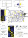
CD4+ T cell help primarily alters the steady-state transcriptome of TEM CD8+ T cells. a, b Volcano plots depicting results of comparative transcriptome analysis of steady-state E7-specific CD8+ TCM cells (a) and TEM cells (b) isolated from spleen at day 100 after primary vaccination with Help or No Help vaccine (n = 3 mice per group). Blue and yellow dots indicate transcripts with significant differential expression (p < 0.01). See also Supplementary Data 1 and 2. c Hierarchical clustering and heat map of the differentially expressed genes in steady-state TEM and TCM cells taken from the Help versus No Help vaccination settings. d Molecules differentially expressed in steady-state TEM cells after Help versus No Help vaccination, as assigned to the indicated functional categories and annotated for subcellular localization by IPA. e Gene Set Enrichment Analysis (GSEA) of published gene sets listing the top 200 up- or downregulated genes in effector CD8 T cells (day 8 after LCMV-Armstrong infection) within the gene expression profile of helped versus helpless TEM cells in steady-state. ES, enrichment score; NES, normalized enrichment score. f Hierarchical clustering and heat map of genes differentially expressed in steady-state TEM cells taken at day 100 after primary vaccination with Help or No Help vaccine, as determined here and genes differentially expressed in primary CTLs taken at day 10 from the same settings, as determined previously. Numbers and adjacent boxes (right panels) indicate Cluster 1, denoting molecules differentially expressed in both helped versus helpless TEM cells and primary CTLs and Cluster 2, denoting molecules only differentially expressed in helped versus helpless TEM cells, as assigned to the indicated functional categories and annotated for subcellular localization by IPA.
Fig. 4
Fig. 4
CD4+ T cell help promotes innate recall function of memory CD8+ T cells. a Experimental set up. CD45.1+ OT-I T cells were adoptively transferred into CD45.2+ recipient mice (n = 5 per group) that 1 day later received OVA-encoding Help or No Help pDNA vaccine. At day 100 after primary vaccination, flow cytometry was performed in blood and CD45.1+ OT-I T cells were flow cytometrically isolated from dLN and spleen and directly analyzed or incubated for 5 h with IL-12 and IL-18 or OVA peptide. Unvaccinated recipient mice were used to isolate naïve OT-I T cells. b Representative flow cytometric plots (left panel) and percentage of IL18-Rα expressing cells within CD45.1+ OT-I cells in indicated organs (right panel). c Representative flow cytometric plots (left panel) and MFI of IL-12Rβ expressed on CD45.1+ OT-I T cells in indicated organs (right panel). dg Primary flow cytometric data (d) and frequencies of IFNγ-producing (e, g) or Granzyme B (GZMB)-producing (f) OT-I T cells in dLN and spleen, as determined after in vitro stimulation with IL12 + IL-18 (D-F) or OVA peptide (SIINFEKL) (g). h Experimental set up. CD45.1+ OT-I T cells were adoptively transferred into CD45.2+ recipient mice (n = 5 per group) that 1 day later received OVA-encoding Help or No Help pDNA vaccine. At day 50 after primary vaccination, mice were rechallenged with E7-encoding Help vaccine and injected with isotype control or neutralizing mAb to IL-12, 4 times, every 3 days. At day 10 after secondary vaccination, total dLN and spleen cells were incubated for 5 h with GolgiPlug (no cytokines or antigens were added). i Frequencies of IFNγ-producing CD45.1+ OT-I T cells isolated from indicated organs. j Representative flow cytometric analysis of cells isolated from spleen. Data are from one experiment representative of two experiments. Error bars indicate SD, *p < 0.05, **p < 0.01, ***p < 0.001 (unpaired two-tailed Student’s t test). Source data are provided as a Source Data file.
Fig. 5
Fig. 5
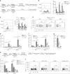
Helped memory CD8+ T cells express the Help signature genes after recall with MHC class I-restricted peptide only. Mice received primary vaccination with Help or No Help vaccine and were recalled at day 50 with No Help vaccine + LPS, as outlined in Fig. 1a. At day 10 of the secondary response, E7-specific CD8+ T cells were flow cytometrically isolated form the spleen (n = 3) and samples were processed for RNAseq. a Volcano plot depicting results of comparative transcriptome analysis of helped versus helpless secondary effectors. Blue and yellow dots indicate transcripts with significant differential expression between the two groups (p < 0.01) See also Supplementary Data 3. b, c Fold change in mRNA expression of a number of signature genes differentially expressed in the Help versus No Help setting of the primary response (1o), as determined before or the secondary response (2o), as determined here. d Hierarchical clustering of genes that were differentially expressed in the Help versus No Help groups in steady-state TEM (Fig. 3), primary (1o) CTLs or secondary (2o) CTLs (a). Certain genes of known or suspected functional or diagnostic relevance are annotated. e Correlation between log2 fold changes in mRNA expression of genes differentially expressed in helped versus helpless TEM cells and primary (1o) CTLs (left panel) or secondary (2o) CTLs (right panel). Error bars indicate SD, *p < 0.05 (unpaired two-tailed Student’s t test). Source data are provided as a Source Data file.
Fig. 6
Fig. 6
Help delivered during priming leaves an epigenetic imprint in steady-state memory cells. Mice (n = 3 per group) received Help or No Help vaccine on days 0, 3, and 6. At day 100, E7-specific memory CD8+ T cells were isolated from the spleens and subjected to ChIPseq analysis with antibodies against H3K4me3 and H3K27me3. a PCA plots depicting results of ChIPseq analysis. b, c Volcano plots depicting differentially modified regions (DMRs) as designated by H3K4me3 (b) or H3K27me3 (c) marks. Blue and red dots indicate DMRs with significant differential modifications (FDR < 0.05). d Molecules differentially modified by H3K4me3 in helped memory E7-specific CD8+ T cells, as assigned to functional categories and annotated for subcellular localization by IPA. e, f Correlation between differential mRNA expression in helped versus non-helped TEM (e) and TCM (f) cells and histone methylation in gene bodies and regions maximally 1 kb upstream from the TSS. Normalized tags of H3K4me3 and H3K27me3 are shown versus average normalized mRNA expression values in helped versus non-helped TEM (e) and TCM (f) cells (described in Fig. 5).

References

    1. Farber DL, Netea MG, Radbruch A, Rajewsky K, Zinkernagel RM. Immunological memory: lessons from the past and a look to the future. Nat. Rev. Immunol. 2016;16:124–128. doi: 10.1038/nri.2016.13. - DOI - PubMed
    1. Lauvau G, Boutet M, Williams TM, Chin SS, Chorro L. Memory CD8+ T cells: innate-like sensors and orchestrators of protection. Trends Immunol. 2016;37:375–385. doi: 10.1016/j.it.2016.04.001. - DOI - PMC - PubMed
    1. Kaech SM, Cui W. Transcriptional control of effector and memory CD8+ T cell differentiation. Nat. Rev. Immunol. 2012;12:749–761. doi: 10.1038/nri3307. - DOI - PMC - PubMed
    1. Joshi NS, Kaech SM. Effector CD8 T cell development: a balancing act between memory cell potential and terminal differentiation. J. Immunol. 2008;180:1309–1315. doi: 10.4049/jimmunol.180.3.1309. - DOI - PubMed
    1. Mueller SN, Gebhardt T, Carbone FR, Heath WR. Memory T Cell Subsets, Migration Patterns, and Tissue Residence. Annu. Rev. Immunol. 2013;31:137–161. doi: 10.1146/annurev-immunol-032712-095954. - DOI - PubMed

Publication types

MeSH terms