

Small Animal MRI
Alcohol Health Res World.
1995.
No abstract available
Keywords: AOD abstinence; AODE (alcohol and other drug effects); animal model; brain damage; magnetic resonance imaging; thiamine deficiency.
Figures
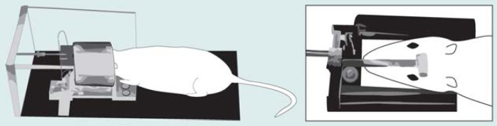
The small animal magnetic resonance imaging apparatus. To ensure precise and reproducible positioning, the rat is lightly anesthetized before being placed in the head brace. The head brace is surrounded by a metal coil that generates the magnetic field and the radiofrequency waves used to create the brain image. The plastic bar (see inset) on the rat’s head is one of the three markers used to align images taken at different times.

Small animal magnetic resonance images showing the effects of thiamine deficiency on the size of fluid-filled brain cavities (ventricles) in the rat. The images are of a lengthwise cross-section of the brain.1 The upper image shows a rat before the start of a thiamine-deficient diet. The lower image is of the same rat after 6 weeks on the diet and shows enlargement of the lateral and third ventricles as well as a loss of fatty tissue and muscles in the rat’s neck region. The crosses and white circles represent three anatomical markers located near the front teeth (incisors), ears, and the top of the skull (bregma) to allow accurate comparison of images taken at different times. SOURCE: Adapted from Pentney, R.J.; Alletto, J.J.; Acara, M.A.; Dlugos, C.A.; and Fiel, R.J. Small animal magnetic resonance imaging: A means of studying the development of structural pathologies in the rat brain. Alcoholism: Clinical and Experimental Research 17(6): 1301–1308,1993.
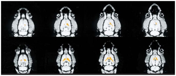
Small animal magnetic resonance images of a rat brain before and after thiamine deficiency. The images represent horizontal cross-sections progressing from the top of the head (left images) to the base of the skull (right images). The images on the top show the rat’s brain before the experiment; those on the bottom were obtained after the rat received a thiamine-deficient diet for 6 weeks. In each of the cross-sections analyzed, the ventricles (indicated in yellow) were larger after thiamine deficiency than before the experiment. SOURCE: Adapted from Pentney. R.J.; Alletto, J.J.; Acara, M.A.; Dlugos, C.A.; and Fiel, R.J. Small animal magnetic resonance imaging: A means of studying the development of structural pathologies in the rat brain. Alcoholism: Clinical and Experimental Research 17(6): 1301–1308.1993.
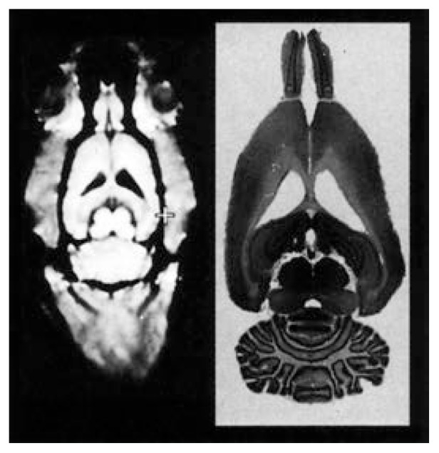
Comparison of a small animal magnetic resonance (SAMRI) image (left) and the histological examination of the corresponding brain slice (right). The SAMRI image shows the brain as well as the surrounding skull. All the brain structures that can be seen in the histological slice are visible in the corresponding image. SOURCE: Adapted from Pentney et al. 1993.
References
-
- Acara M, Bilotta J, Pazik M, Goldfarb D. Transport and metabolism of thiamine by the chicken and rat kidney. Effect of ethanol. (Abstract) Pharmacologist. 1983;25:345.
-
- Ballon D, Graham MC, Miodownik S. Doubly tuned solenoidal resonators for small animal imaging and spectroscopy at 1.5 Tesla. Magnetic Resonance Imaging. 1989;7:155–162. - PubMed
-
- Butterworth R. Effects of thiamine deficiency on brain metabolism: Implications for the pathogenesis of the Wernicke-Korsakoff syndrome. Alcohol and Alcoholism. 1989;24:271–279. - PubMed
-
- Butterworth R, Kril JJ, Harper CG. Thiamine-dependent enzyme changes in the brains of alcoholics: Relationship to the Wernicke-Korsakoff syndrome. Alcoholism: Clinical and Experimental Research. 1993;17:1084–1088. - PubMed
-
- Chick JD, Smith MA, Engleman HM, Kean DM, Mander A, Douglas RHB, Best JK. Magnetic resonance imaging of the brain of alcoholics: Cerebral atrophy, lifetime alcohol consumption, and cognitive deficits. Alcoholism: Clinical and Experimental Research. 1989;13:512–518. - PubMed
LinkOut - more resources
Full Text Sources